유방암 치료결정에 도움이 될 수 있는 영상검사
2021.03.15 · amschool
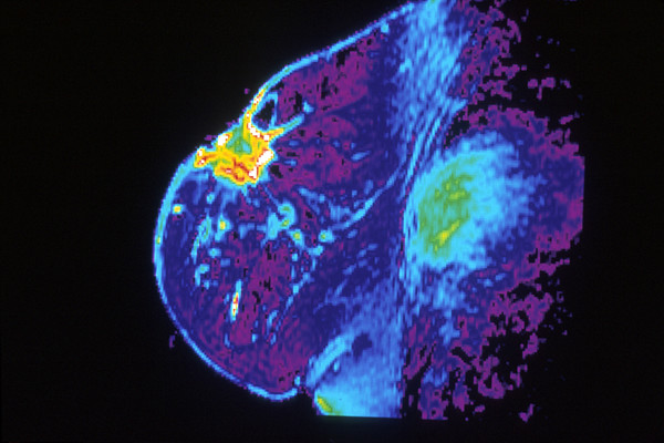
새로운 영상검사가 일부 유방암 환자들의 치료 결정을 하는 데 도움이 될 수 있다고 국립암연구소가 후원하는 소규모 임상 연구 결과가 시사한다. 이번 실험은 타목시펜(상표명 : 놀바덱스)이나 레트로졸(상표명 : 페마라)처럼 호르몬 차단 요법에 반응해야 하는 종양을 찾아내기 위한 것으로 나타났다.
영상접근법은 유방암이 에스트로겐 수용체(ER) 양성인 사람들을 대상으로 한 것으로, 종양 세포에는 에스트로겐 호르몬에 대한 수용체가 풍부하게 들어 있으며 이러한 암의 성장은 에스트로겐에 의해 촉진된다는 것을 의미한다. 여성 유방암의 약 70-80%, 남성 유방암의 약 90%가 ER 양성이다.
호르몬 치료는 ER 양성 유방암 치료의 주축이다. 내분비 치료법으로도 알려진 이 약물들은 에스트로겐의 종양을 굶기거나 에스트로겐이 수용체에 부착되거나 결합되는 것을 막음으로써 종양 성장을 늦추거나 중단시킨다.
하지만 ER 양성 종양을 가진 모든 사람이 호르몬 치료에 반응하는 것은 아니며, 많은 사람들이 결국 약물에 내성을 갖게 될 것이다.
이 새로운 영상 검사는 종양 내의 에스트로겐 수용체가 활동적이어서 에스트로겐에 반응하는지 여부를 보여줌으로써 어떤 환자들이 이러한 내성이 있는 종양을 가지고 있는지를 알아내는데 도움을 줄 수 있다. 연구에서, 종양이 활동적인 에스트로겐 수용체를 가지고 있는 모든 환자들은 호르몬 치료를 받으면 그들의 질병이 개선되거나 안정되어 있는 것을 보았다. 이와는 대조적으로, 종양에 활성 에스트로겐 수용체가 부족한 모든 여성들에게서 이 질병이 진행되었다고 연구원들은 2월 2일 “네이처 커뮤니케이션”에 보고했다.
이 새로운 연구에 참여하지 않은 국립암연구소 암 연구 센터의 여성 악성 종양학과의 스탠리 립코위츠 박사는 "의사들이 가장 도움이 될 가능성이 높은 환자들에게 호르몬 요법의 사용을 맞춤화할 수 있도록 허용함으로써, 영상 검사는 종양학자들에게 잠재적으로 귀중한 수단을 제공한다."고 말했다.
더 규모가 큰 연구에서 그 결과가 확인되어야 하지만, 이 시험은 "흥미롭고" "기대가 된다"고 밴더빌트 대학 의료센터의 의료 종양학자이자 유방암 전문가인 로라 케네디는 말했다.
이번 검사를 통해 ER 양성 유방암 환자들이 수개월 동안 병이 진행될 수 있는 비효과적인 치료를 피하는 데 도움이 될 수 있을 것이라고 워싱턴 의과대학의 연구리더인 파로크 데다시티 박사가 말했다.
에스트로겐 수용체는 종양 속에 있지만, 그것들은 활동적인가?
유방암 세포에 에스트로겐 수용체가 들어 있는지 알아보기 위해 의사들은 현재 외과적으로 채취한 종양조직 샘플이나 침 생검(생체검사 혹은 조직검사)을 하고 있다.
치료 후 재발하거나 신체의 다른 부위로 퍼진(전이) 암인 진행성 ER 양성 유방암 환자들은 대개 호르몬 치료를 시작한 다음 암이 치료에 반응하는지 관찰한다.
그러나 현재 실험은 종양 세포의 에스트로겐 수용체가 종양의 성장을 촉진하고 있는지, 따라서 호르몬 치료에 반응할 가능성이 있는지에 대한 어떠한 정보도 제공하지 않는다.
에스트로겐 수용체가 작용하는 정상 세포뿐만 아니라 종양 세포에서도 에스트로겐은 에스트로겐 수용체와 결합해 작동시킨다. 활성화된 에스트로겐 수용체는 많은 세포 과정에 영향을 미친다. 유방암 세포가 에스트로겐에 반응하는 한 가지 방법은 다른 종류의 호르몬 수용체인 프로게스테론 수용체를 더 많이 만드는 것이다.
이 새로운 테스트는 환자들이 짧은 시간 동안 에스트로겐을 투여 받은 후 종양 내 프로게스테론 수용체 수치의 변화를 찾는다. 종양 세포의 에스트로겐 수용체가 에스트로겐에 반응한다면 프로게스테론 수용체 수치가 올라가야 한다.
이 테스트는 프로게스테론 수용체에 특별히 결합하는 FFNP라고 불리는 방사능 추적기를 사용하는 PET 스캔을 포함한다. 프로게스테론 수용체가 더 많이 존재할 때, 종양 세포가 차지하는 FFNP의 양은 더 크고 PET 스캔에서 FFNP의 신호는 더 강해진다.
FFNP를 사용하여 종양을 밝히다
이 작은 임상실험을 위해, 데다시티 박사 팀은 43명의 폐경 후 ER 양성 유방암을 가진 여성들을 연구했습니다. 그들은 PET-CT 스캔에서 FFNP를 화상 작용제로 사용했는데, 이 스캔은 PET 스캔을 X선의 형태로 사용하는 영상촬영 방법인 CT 스캔과 결합한다. PET-CT 스캔은 PET 스캔만으로 얻을 수 있는 것보다 FFNP로 "밝아진" 종양의 해부학적 위치에 대한 더 정확한 정보를 제공한다.
환자들은 처음에 FFNP로 PET-CT 스캔을 받았다. 그르고 나서 그들은 24시간 동안 3회분의 에스트로겐을 투여 받았고 FFNP를 이용한 영상 테스트는 반복되었다. 24시간 에스트로겐 "도전" 전후의 PET-CT 스캔은 종양에 의해 FFNP의 양이 달라지는 것을 관찰하기 위해 비교되었다.
스캔이 완료된 후, 모든 환자들은 종양학자들의 권유로 다양한 종류의 호르몬 치료를 받았다. 연구팀은 최소 6개월 동안 모든 환자를 추적하고 치료에 대한 반응을 관찰했다.
유방암은 43명의 환자 중 15명(비 응답자)이 호르몬 치료를 시작한 지 6개월 만에 진행됐으며, 나머지 28명(응답자)은 안정적이거나 호전된 상태를 유지했다. 이 28명의 여성들은 종양이 치료에 반응하지 않는 15명보다 더 오래 살았다.
그런 다음 연구원들은 응답자와 비 응답자의 FFNP PET-CT 스캔을 비교했다
첫 번째 PET-CT 스캔에서 환자의 종양이 받아들인 FFNP의 양은 응답자와 비 응답자 간에 차이가 없었다. 그러나 첫 번째와 두 번째 PET-CT 스캔 사이의 변화는 효과가 있었다. 에스트로겐 투여 후, 종양이 받아들인 FFNP의 양은 나중에 호르몬 치료에 반응한 28명의 환자들 모두에서 증가했다. 이와는 대조적으로, 종양의 FFNP 흡수는 호르몬 치료에 반응하지 않는 15명의 환자들 모두에서, 대부분의 경우, 혹은 약간만 증가했다.
일부 환자들은 하루 동안의 에스트로겐 “도전”에 이어 단기적인 부작용을 경험했다. 대부분 경미한 부작용으로는 설사, 메스꺼움, 구토, 근골격계 통증 등이 있었다. FFNP가 주어진 후 부작용은 나타나지 않았다.
임상 치료에 잘 통합될 수 있는 테스트
데다슈티 박사는 "이것은 단일 의료센터에서 시행된 소규모 임상실험으로, 유방암에 걸린 사람들에게 일상적으로 이 검사를 이용하려면 더 큰 규모의 여러 센터 실험에서 반복될 필요가 있다"고 말했다.
만약 연구 결과가 확인된다면, 새로운 에스트로겐 “도전” 테스트는 에스트로겐 수용체가 기능적인지 아닌지를 보여주는 것 이상의 이점을 조직 생체검사에 제공할 것이라고 그녀는 말했다. 데다슈티 박사는 "생체검사는 보통 종양조직의 작은 부분만을 채취하는 반면 FFNP 영상검사제는 의사들이 종양 전체를 볼 수 있게 하고 생체검사에서는 쉽게 볼 수 없는 암 병변을 포함한 신체의 모든 암 병변을 한 번에 볼 수 있게 한다."고 설명했다.
"체내의 모든 종양에서 에스트로겐 수용체 기능을 가시화하는 능력은 상당한 이점"이라고 이번 연구에 참여하지 않은 NCI의 암 화상 프로그램 담당 부국장인 자넷 어리 박사가 말했다. 왜냐하면 에스트로겐 수용체의 존재와 아마도 수용체가 에스트로겐에 반응하는지 아닌지는 개인 내의 전이성 종양들 사이에서 상당히 다양할 수 있기 때문에 치료 결정에 영향을 미칠 수 있기 때문이다. 그래서 이런 테스트를 하는 것은 판도를 바꾸는 것일 수 있다.
"비효과적인 치료의 비용은 어마어마하다"고 어리 박사는 말했다. 재정적인 비용뿐만 아니라 인명의 잠재적인 손실이나 생명의 단축의 측면에서도 그러하다.
"이 테스트는 실제로 매우 잘 통합될 것이다. 우리는 종종 전이성 유방암 환자들에게서 PET 스캔을 한다"고 케네디 박사는 말했다. "우리는 이러한 환자들의 [ER-양성] 종양들이 궁극적으로 호르몬 치료에 내성을 갖게 될 것이라는 것을 알고 있다. 따라서 만약 누군가의 암이 빨리 진행된다면, 어떤 환자들은 더 빨리 화학요법으로 전환되어야 한다는 것을 알려주는 [하루] 검사를 받는 것이 도움이 될 것이다."
립코위츠 박사는 "이번 실험은 또 다른 종류의 호르몬 요법에 대한 내성이 생긴 후에도 여전히 반응할 수 있는 환자들을 가려내는 데 도움이 될 것"이라고 말했다.
케네디 박사는 또한 이번 연구에는 이전에 치료를 받지 않은 환자, 호르몬 치료만을 받은 환자, 호르몬 치료와 병행하여 다른 치료를 받은 환자 등 광범위한 환자가 포함된다고 언급했다.
그 결과, 그녀는 더 규모가 클 뿐 아니라 암으로 유사한 치료를 받는 환자 집단을 포함하는 연구에서 이 검사가 얼마나 잘 수행되는지 보는 것이 중요할 것이라고 말했다. 보다 동종 환자 군을 연구하면 호르몬 요법민감성 환자 대 호르몬요법 내성 종양 환자를 구별할 수 있는 능력의 한계를 보다 효과적으로 드러낼 수 있다고 그녀는 말했다.
또한 에스트로겐의 순환량이 자연적으로 높기 때문에 폐경 전 여성들에게 에스트로겐 도전이 가능할지 여부는 분명하지 않다고 립코위츠 박사는 지적했다.
이 시험의 또 다른 한계 중 하나는 정상적인 간이 FFNP를 다량 받아들이기 때문에 간으로만 전이된 유방암 환자들에게는 사용할 수 없다는 점이라고 연구진은 지적한다.
2021년 3월 11일 : 국립암연구소
Imaging Test Could Help Guide Breast Cancer Treatment Decisions
March 11, 2021, by NCI Staff
A new imaging test could help guide treatment decisions for some people with advanced breast cancer, results of a small NCI-funded clinical study suggest. The test is designed to identify tumors that should respond to hormone-blocking therapies like tamoxifen (Nolvadex) or letrozole (Femara), the study showed.
The imaging approach is for people whose breast cancer is estrogen-receptor (ER) positive, which means that their tumor cells contain an abundance of receptors for the hormone estrogen and the growth of these cancers is fueled by estrogen. About 70%–80% of breast cancers in women and 90% in men are ER positive.
Hormone therapies are a mainstay of treatment for ER-positive breast cancer. Also known as endocrine therapies, these drugs slow or stop tumor growth either by starving the tumors of estrogen or blocking estrogen from attaching, or binding, to the receptor.
But not everyone with ER-positive tumors will respond to hormone therapy, and many will eventually become resistant to the drugs.
The new imaging test may help pinpoint which patients have these resistant tumors by showing whether the estrogen receptors in tumors are active and responsive to estrogen. In the study, all patients whose tumors had active estrogen receptors saw their disease improve or remain stableExit Disclaimer when given hormone therapy. By contrast, the disease progressed in all the women whose tumors lacked active estrogen receptors, researchers reported February 2 in Nature Communications.
By allowing doctors to tailor the use of hormone therapy to those patients who are most likely to benefit, the imaging test offers a potentially valuable tool for oncologists, said Stanley Lipkowitz, M.D., Ph.D., of the Women’s Malignancies Branch in NCI’s Center for Cancer Research, who was not involved in the new study.
Although the results need to be confirmed in a larger study, they are “exciting” and “promising,” said Laura Kennedy, M.D., Ph.D., a medical oncologist and breast cancer specialist at Vanderbilt University Medical Center, who also was not involved with the study.
The test could help patients with ER-positive breast cancer avoid months of ineffective treatment during which their disease may progress, said study leader Farrokh Dehdashti, M.D., of the Washington University School of Medicine in St. Louis.
Estrogen Receptors Are in the Tumors, but Are They Active?
To find out whether breast cancer cells contain estrogen receptors, doctors currently test samples of tumor tissue that have been removed surgically or with a needle biopsy.
Patients with advanced ER-positive breast cancer—cancer that has come back after treatment or spread (metastasized) to other parts of the body—usually are started on hormone therapy and then monitored to see if their cancer responds to treatment.
However, current tests don’t provide any information on whether the estrogen receptors in tumor cells are driving the tumor’s growth and, thus, are likely to respond to hormone therapy.
In tumor cells as well as normal cells with working estrogen receptors, estrogen binds to the estrogen receptor and turns it on. The activated estrogen receptor then influences many cellular processes. One way that breast cancer cells respond to estrogen is by making more of another type of hormone receptor, the progesterone receptor.
The new test looks for changes in the levels of progesterone receptors in tumors after patients receive estrogen for a short time. If estrogen receptors in the tumor cells are responsive to estrogen, then progesterone receptor levels should go up.
The test involves a PET scan that uses a radioactive tracer called FFNP, which binds specifically to progesterone receptors. When more progesterone receptors are present, the amount of FFNP taken up by tumor cells is greater and the signal from FFNP on a PET scan is stronger.
Using FFNP to “Light Up” Tumors
For this small clinical trial, Dr. Dehdashti’s team studied 43 postmenopausal women with advanced ER-positive breast cancer. They used FFNP as an imaging agent in PET-CT scans, which combine PET scans with CT scans, an imaging method that uses a form of x-rays. PET-CT scans provide more precise information about the anatomical location of tumors that “light up” with FFNP than a PET scan alone can provide.
Patients first underwent PET-CT scans with FFNP. They were then given three doses of estrogen over a 24-hour period, and the imaging test with FFNP was repeated. PET-CT scans before and after the 24-hour estrogen “challenge” were compared to look for changes in the amount of FFNP taken up by tumors.
After the scans were completed, all of the patients received various types of hormone therapy, as recommended by their oncologists. The team followed all patients for a minimum of 6 months and monitored their responses to treatment.
Breast cancer progressed in 15 of the 43 patients (nonresponders) within 6 months of starting hormone therapy, and the disease remained stable or improved in the other 28 patients (responders). These 28 women lived longer than the 15 whose tumors did not respond to therapy.
The researchers then compared the FFNP PET-CT scans of responders and nonresponders.
The amount of FFNP taken up by patients’ tumors on the first PET-CT scans did not differ between responders and nonresponders. But the change between the first and second PET-CT scans did. After the estrogen challenge, the amount of FFNP taken up by tumors went up in all 28 patients whose disease later responded to hormone therapy. By contrast, tumor uptake of FFNP went down in most cases, or up only modestly, in all 15 patients whose disease did not respond to hormone therapy.
Some patients experienced short-term side effects following the one-day estrogen challenge. These side effects, most of which were mild, included diarrhea, nausea and vomiting, and musculoskeletal pain. No side effects were seen after FFNP was given.
Test Would Integrate Well into Clinical Care
This was a small clinical trial done at a single medical center and needs to be repeated in a larger multicenter trial before the test can be used routinely in people with advanced breast cancer, Dr. Dehdashti said.
If the findings are confirmed, she said, the new estrogen challenge test would offer advantages over tissue biopsies beyond showing whether estrogen receptors are functional. Whereas a biopsy usually samples only a small piece of tumor tissue, the FFNP imaging agent lets doctors see the entire tumor as well as showing all of the cancer lesions in the body at one time, including those that can’t be biopsied readily, Dr. Dehdashti explained.
The ability to visualize estrogen receptor function in all the tumors in the body is a significant advantage, said Janet Eary, M.D., associate director of NCI’s Cancer Imaging Program, who was not involved with the study. That’s because the presence of estrogen receptors—and presumably whether the receptors are responsive to estrogen or not—can vary quite a bit among metastatic tumors within an individual, which could also influence treatment decisions.
So, having a test like this could be a game changer.
“The cost of ineffective treatment is enormous,” not only in terms of financial cost but also the potential loss or shortening of human life, Dr. Eary said.
“This test would integrate really well into practice, where we often do PET scans in patients with metastatic breast cancer,” Dr. Kennedy said. “We know that those patients’ [ER-positive] tumors ultimately will become resistant to hormone therapy, and so if someone’s cancer is progressing quickly, it might be helpful to have a [one day] test to let you know that some patients should be switched to chemotherapy sooner.”
The test also could help identify patients who may still respond to another type of hormone therapy after their disease becomes resistant to, and progresses on, the first hormone therapy given, Dr. Lipkowitz said.
Dr. Kennedy also noted that the study included a broad mix of patients, including some who had not previously been treated, some who received hormone therapy alone, and others who received other treatments in combination with hormone therapy.
As a result, she said it will be important to see how well the test performs in a study that is not only larger but also involves a group of patients who receive similar treatments for their cancer. Studying more homogeneous groups of patients could more effectively reveal limitations in the test’s ability to distinguish between patients with hormone therapy–sensitive versus –resistant tumors, she said.
It is also not clear if the estrogen challenge would be feasible in premenopausal women because their circulating estrogen levels are naturally high, Dr. Lipkowitz noted.
One other limitation of the test, the study investigators note, is that it cannot be used in patients with breast cancer that has spread only to the liver because normal liver takes up large amounts of
암스쿨에 게재된 기사는 미국국립암연구소(NCI), 미국암협회(ACS), 국립암센터(NCC), 일본국립암연구소(NCCJ), 엠디앤더슨암센터(MD Anderson Cancer Center) 등 검증된 기관의 검증된 자료를 토대로 작성되었습니다.
이 글은 2021년 3월 작성 시점의 정보입니다. 치료 기준·급여 정보는 바뀌었을 수 있으니, 진료와 치료 결정은 반드시 담당 의료진과 상의하세요.