종양의 등급이란 무엇인가?
2020.10.27 · 전영조 전문위원

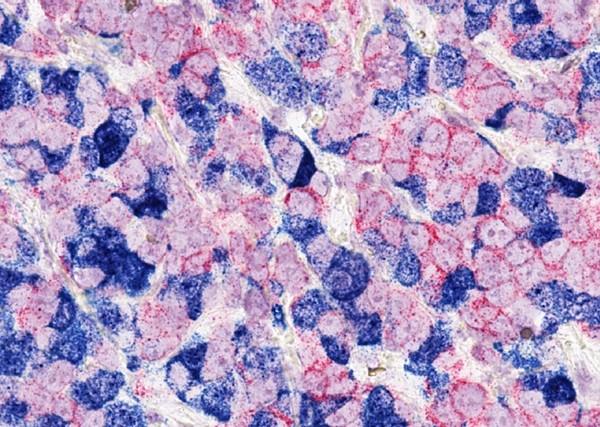
종양등급은 종양세포와 종양조직이 현미경 검사에서 얼마나 비정상적으로 보이는가를 기준으로 종양을 기술한 것이다. 종양이 얼마나 빨리 자라고 퍼질지 보여주는 지표다. 종양의 세포와 종양의 조직이 정상 세포와 조직의 세포에 가까우면 종양을 '잘 분화된' 것으로 부른다. 이러한 종양들은 비정상적으로 보이는 세포를 가지고 있고 정상적인 조직 구조가 결여될 수 있는 "미분화" 또는 "분화 불량"인 종양보다 느린 속도로 성장하고 퍼지는 경향이 있다. 현미경의 외관상의 이것들과 다른 차이점을 근거로, 의사들은 대부분의 암에 수치적인 "등급"을 부여한다. 종양 등급을 결정하는 데 사용되는 요인은 여러 종류의 암에 따라 다를 수 있다.
종양 등급은 암의 병기와 같지 않다. 암의 병기는 원발 (1차) 종양의 크기 및/또는 도달 범위와 체내에 암세포가 퍼졌는지 여부를 가리킨다. 암의 병기는 1차 종양의 위치, 종양 크기, 국부 림프절 관여(주변 림프절에 암이 전이됨), 존재하는 종양의 수 등의 요인에 기초한다. 암 발생에 대한 자세한 내용은 ‘병기 측정’ 페이지에서 확인할 수 있다.
종양 등급은 어떻게 결정되는가?
만약 종양이 악성인 것으로 의심되면 의사는 생검이라는 시술 중에 종양의 전부 또는 일부를 떼어낸다. 이어 병리학자(현미경으로 세포와 조직을 연구해 질병을 식별하는 의사)가 생체 조직을 검사해 종양이 양성인지 악성인지 판별한다. 병리학자는 또한 종양의 등급을 결정하고 종양의 다른 특징을 확인한다. 국립암연구소 자료표의 병리학 보고서는 생검이나 기타 수술 중에 적출한 조직의 시각 및 현미경 검사에 관한 병리학자의 보고서에서 찾을 수 있는 정보의 종류이다.
종양 등급은 어떻게 분류되는가?
등급 시스템은 암의 종류에 따라 다르다. 일반적으로 종양은 이상의 양에 따라 1, 2, 3, 4로 등급이 매겨진다. 1급 종양에서는 종양 세포와 종양 조직의 조직이 정상 가까이 나타난다. 이러한 종양들은 천천히 자라고 퍼지는 경향이 있다. 대조적으로 3급과 4급 종양의 세포와 조직은 정상 세포와 조직처럼 보이지 않는다. 3급과 4급 종양은 급성장하고 등급이 낮은 종양보다 더 빨리 퍼지는 경향이 있다.
종양 종류에 대한 등급제를 지정하지 않은 경우, 일반적으로 다음과 같은 시스템이 사용된다(1)
▲ GX : 등급은 평가할 수 없다(미정 등급)
▲ G1 : 잘 분화된(낮은 등급)
▲ G2 : 적당히 분화된(중간 등급)
▲ G3 : 분화 불량(높은 등급)
▲ G4: 미분화(높은 등급)
암 유형별 고유의 등급 체계는 어떤 것이 있는가?
유방암과 전립선암은 자체 등급제가 있는 암의 가장 흔한 유형이다. 유방암. 의사들은 유방암에 대해 노팅엄 등급 체계(스카프-블룸-리차드슨 등급 체계의 엘스턴-엘리스 수정본이라고도 한다)를 가장 많이 사용한다. 이 시스템은 다음과 같은 특징에 따라 유방 종양을 등급화 한다.
▲ 세관 형성 : 종양 조직 중 정상 유관 구조를 가진 조직 수
▲ 핵의 등급 : 종양세포 내 핵의 크기와 형태 평가
▲ 유사 분열률 : 몇 개의 분열세포가 존재하는지, 이것은 종양세포가 얼마나 빨리 자라고 분열하고 있는지를 나타내는 척도이다.
각각의 카테고리는 1과 3 사이의 점수를 받는다. "1"의 점수는 세포와 종양 조직이 정상 세포와 조직과 가장 닮았다는 것을 의미하고, 3의 점수는 세포와 조직이 가장 비정상적으로 보인다는 것을 의미한다. 그런 다음 세 가지 카테고리의 점수를 합산하여 총점 3~9를 산출한다. 세 가지 등급이 가능하다.
▲ 총점수 = 3–5: G1 (낮은 등급 또는 잘 차별화된 등급)
▲ 총점수 = 6-7: G2(중간등급 또는 적당히 분화된 등급)
▲ 총점수 = 8~9: G3(등급이 높거나 분화 불량의 등급)
전립선암. 글리슨 채점 시스템은 전립선암 등급을 매기기 위해 사용된다. 글리슨 점수는 전립선에서 채취한 조직검사 검체(표본)를 바탕으로 한다. 병리학자는 종양 조직이 정상 전립선 조직과 얼마나 비슷하게 보이는지 샘플을 검사한다. 조직의 원발 패턴과 2차 패턴을 모두 확인한다. 원발(1차) 패턴은 종양에서 볼 수 있는 가장 일반적인 조직 패턴을 나타내며, 2차 패턴은 다음으로 가장 일반적인 패턴을 나타낸다. 각각의 패턴에는 1등급부터 5등급까지의 등급이 부여되며, 1등급은 정상 전립선 조직과 가장 유사해 보이고 5등급은 비정상적으로 보인다. 그리고 두 등급이 추가되어 글리슨 점수가 된다. 미국 암 공동 위원회는 글리슨 점수를 다음 범주로 분류할 것을 권고한다.
▲ 글리슨 X: 글리슨 점수를 확인할 수 없다.
▲ 글리슨 2-6: 종양 조직은 잘 분화되었다.
▲ 글리슨 7: 종양 조직이 적당히 분화되었다.
▲ 글리슨 8-10: 종양 조직이 분화 불량이거나 미분화 상태이다..
종양 등급이 환자의 치료 선택에 어떤 영향을 미치는가?
의사들은 종양 등급과 병기, 환자의 나이, 일반 건강 등의 다른 요인을 이용하여 치료 계획을 수립하고 환자의 예후(질병의 예상 가능한 결과나 과정, 회복이나 재발의 가능성)를 결정한다. 일반적으로 등급이 낮으면 예후가 더 좋다는 뜻이다. 더 높은 등급의 암은 더 빨리 자라고 퍼질 수 있으며 즉각적이거나 더 공격적인 치료가 필요할 수 있다.
연부조직 육종과 원발 뇌종양, 유방 및 전립선암 등 특정 암의 경우 치료 계획과 환자의 예후 결정 시 종양 등급의 중요성이 더 크다. 환자들은 종양 등급에 대한 더 많은 정보와 그것이 그들의 치료와 예후와 어떻게 관련이 있는지에 대해 의사와 상의해야 한다.
검토 : 2013년 5월 3일
Tumor Grade
Reviewed: May 3, 2013
On This Page
• What is tumor grade?
• How is tumor grade determined?
• How are tumor grades classified?
• What are some of the cancer type-specific grading systems?
• How does tumor grade affect a patient’s treatment options?
What is tumor grade?
Tumor grade is the description of a tumor based on how abnormal the tumor cells and the tumor tissue look under a microscope. It is an indicator of how quickly a tumor is likely to grow and spread. If the cells of the tumor and the organization of the tumor’s tissue are close to those of normal cells and tissue, the tumor is called “well-differentiated .” These tumors tend to grow and spread at a slower rate than tumors that are “undifferentiated” or “poorly differentiated,” which have abnormal-looking cells and may lack normal tissue structures. Based on these and other differences in microscopic appearance, doctors assign a numerical “grade” to most cancers. The factors used to determine tumor grade can vary between different types of cancer.
Tumor grade is not the same as the stage of a cancer. Cancer stage refers to the size and/or extent (reach) of the original (primary) tumor and whether or not cancer cells have spread in the body. Cancer stage is based on factors such as the location of the primary tumor, tumor size, regional lymph node involvement (the spread of cancer to nearby lymph nodes), and the number of tumors present. More information about cancer staging is available on the Staging page.
How is tumor grade determined?
If a tumor is suspected to be malignant, a doctor removes all or part of it during a procedure called a biopsy. A pathologist (a doctor who identifies diseases by studying cells and tissues under a microscope) then examines the biopsied tissue to determine whether the tumor is benign or malignant. The pathologist also determines the tumor’s grade and identifies other characteristics of the tumor. The NCI fact sheet Pathology Reports describes the type of information that can be found in a pathologist’s report about the visual and microscopic examination of tissue removed during a biopsy or other surgery.
How are tumor grades classified?
Grading systems differ depending on the type of cancer. In general, tumors are graded as 1, 2, 3, or 4, depending on the amount of abnormality. In Grade 1 tumors, the tumor cells and the organization of the tumor tissue appear close to normal. These tumors tend to grow and spread slowly. In contrast, the cells and tissue of Grade 3 and Grade 4 tumors do not look like normal cells and tissue. Grade 3 and Grade 4 tumors tend to grow rapidly and spread faster than tumors with a lower grade.
If a grading system for a tumor type is not specified, the following system is generally used (1):
•GX: Grade cannot be assessed (undetermined grade)
•G1: Well differentiated (low grade)
•G2: Moderately differentiated (intermediate grade)
•G3: Poorly differentiated (high grade)
•G4: Undifferentiated (high grade)
What are some of the cancer type-specific grading systems?
Breast and prostate cancers are the most common types of cancer that have their own grading systems.
Breast cancer. Doctors most often use the Nottingham grading system (also called the Elston-Ellis modification of the Scarff-Bloom-Richardson grading system) for breast cancer (1). This system grades breast tumors based on the following features:
•Tubule formation: how much of the tumor tissue has normal breast (milk) duct structures
•Nuclear grade: an evaluation of the size and shape of the nucleus in the tumor cells
•Mitotic rate: how many dividing cells are present, which is a measure of how fast the tumor cells are growing and dividing
Each of the categories gets a score between 1 and 3; a score of “1” means the cells and tumor tissue look the most like normal cells and tissue, and a score of “3” means the cells and tissue look the most abnormal. The scores for the three categories are then added, yielding a total score of 3 to 9. Three grades are possible:
•Total score = 3–5: G1 (Low grade or well differentiated)
•Total score = 6–7: G2 (Intermediate grade or moderately differentiated)
•Total score = 8–9: G3 (High grade or poorly differentiated)
Prostate cancer. The Gleason scoring system is used to grade prostate cancer (1). The Gleason score is based on biopsy samples taken from the prostate. The pathologist checks the samples to see how similar the tumor tissue looks to normal prostate tissue. Both a primary and a secondary pattern of tissue organization are identified. The primary pattern represents the most common tissue pattern seen in the tumor, and the secondary pattern represents the next most common pattern. Each pattern is given a grade from 1 to 5, with 1 looking the most like normal prostate tissue and 5 looking the most abnormal. The two grades are then added to give a Gleason score. The American Joint Committee on Cancer recommends grouping Gleason scores into the following categories (1):
•Gleason X: Gleason score cannot be determined
•Gleason 2–6: The tumor tissue is well differentiated
•Gleason 7: The tumor tissue is moderately differentiated
•Gleason 8–10: The tumor tissue is poorly differentiated or undifferentiated
How does tumor grade affect a patient’s treatment options?
Doctors use tumor grade and other factors, such as cancer stage and a patient’s age and general health, to develop a treatment plan and to determine a patient’s prognosis (the likely outcome or course of a disease; the chance of recovery or recurrence). Generally, a lower grade indicates a better prognosis. A higher-grade cancer may grow and spread more quickly and may require immediate or more aggressive treatment.
The importance of tumor grade in planning treatment and determining a patient’s prognosis is greater for certain types of cancer, such as soft tissue sarcoma, primary brain tumors, and breast and prostate cancer.
Patients should talk with their doctor for more information about tumor grade and how it relates to their treatment and prognosis.
암스쿨에 게재된 기사는 미국국립암연구소(NCI), 미국암협회(ACS), 국립암센터(NCC), 일본국립암연구소(NCCJ), 엠디앤더슨암센터(MD Anderson Cancer Center) 등 검증된 기관의 검증된 자료를 토대로 작성되었습니다.
이 글은 2020년 10월 작성 시점의 정보입니다. 치료 기준·급여 정보는 바뀌었을 수 있으니, 진료와 치료 결정은 반드시 담당 의료진과 상의하세요.