감시 림프절의 생체검사를 하는 이유는 무엇인가?
2020.08.10 · 전영조 전문위원
림프절이란 무엇인가?
림프절은 몸의 림프계에 속하는 작고 둥근 기관이다. 림프계는 면역체계의 일부분이다. 그것은 감염을 억제하는 백혈구를 운반하는 투명한 액체인 림프를 포함하는 혈관과 장기의 네트워크와 인체의 세포와 조직에서 나오는 액체와 노폐물들로 구성되어 있다. 암에 걸린 사람에게 림프는 주 종양에서 분리된 암세포를 운반할 수도 있다.
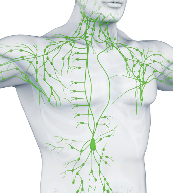
림프는 림프절을 통해 여과되는데, 림프절은 몸 전체에서 광범위하게 발견되며 림프관에 의해 서로 연결되어 있다. 림프절 그룹은 목, 겨드랑이, 가슴, 복부, 사타구니에 위치한다. 림프절은 백혈구(B 림프구와 T 림프구)와 다른 종류의 면역 체계 세포를 포함하고 있다. 림프절은 박테리아와 바이러스뿐만 아니라 일부 손상되고 비정상적인 세포들을 가두어 면역체계가 질병과 싸우도록 돕는다. 많은 종류의 암이 림프계를 통해 퍼지고 있으며, 이러한 암의 가장 초기 확산 부위 중 하나는 근처의 림프절이다.
감시 림프절이란 무엇인가?
감시 림프절은 암세포가 1차 종양에서 가장 잘 퍼질 가능성이 있는 첫 번째 림프절이라고 정의된다. 때때로, 하나 이상의 감시 림프절이 있을 수 있다.
감시 림프절 생체검사란?
감시 림프절 생체검사(SLNB)는 감시 림프절을 식별, 제거, 검사하여 암세포가 존재하는지 여부를 판단하는 절차다. 그것은 이미 암 진단을 받은 사람들에게 사용된다. SLNB 음성 결과는 암이 아직 인근 림프절이나 다른 장기로 전이되지 않았음을 시사한다.
SLNB 양성 결과는 감시 림프절에 암이 존재하며, 암이 근처의 다른 림프절(지역 림프절이라고 함) 및 다른 장기로 전이되었을 수 있음을 나타낸다. 이 정보는 의사가 암의 병기를 결정하고 적절한 치료 계획을 수립하는 데 도움을 줄 수 있다.
SLNB 중에 발생하는 현상
첫째, 감시 림프절이 위치해야 한다. 이를 위해 외과의사는 종양 근처에 방사성 물질, 청색 염료, 또는 둘 다 주사한다. 그런 다음, 외과의사는 방사능 물질이 함유된 림프절을 감지하거나 파란 염료로 얼룩진 림프절을 찾는 장치를 사용한다. 일단 감시 림프절이 파악되면 외과의사는 위쪽의 피부에 작은 절개(약 1/2인치)를 하고 림프절을 제거한다.
그런 다음 병리학자에 의해 감시 림프절에 암세포의 존재 여부를 검사한다. 암이 발견될 경우, 외과의사는 동일한 조직검사 절차 또는 후속 수술 절차 중에 추가로 림프절을 제거할 수 있다. SLNB는 통원으로 절제하거나 병원에 잠시 입원해야 할 수 있다.
SLNB는 보통 원발 종양을 제거하는 동시에 이루어진다. 어떤 경우에는 종양을 제거하기 전이나 후에(임프관이 얼마나 막혔는지에 따라) 시술도 할 수 있다.
SLNB의 이점은 무엇인가?
SNLB는 의사들이 암의 병기 측정하는 것을 돕고, 종양 세포가 신체의 다른 부분으로 퍼지는 능력을 발달시켰을 위험을 추정하도록 돕는다. 만약 감시 림프절에 암이 음성이라면, 환자는 더 광범위한 림프절 수술을 피할 수 있을 것이고, 많은 림프절이 제거되는 것과 관련된 잠재적 합병증을 줄일 수 있을 것이다.
SLNB의 있을 수 있는 해악은 무엇인가?
림프절 절제는 대개 부작용, 특히 림프절과 같은 심각한 부작용과 관련이 적지만 SLNB를 포함한 모든 림프절 절제 수술은 림프종 같은 해로운 부작용을 일으킬 수 있다. 잠재적 부작용은 다음과 같다. :
▲ 림프종 또는 조직 부종. 림프절 수술 중에는 감시 림프절이나 림프절의 그룹으로 이어지는 림프관이 절단된다. 이로 인해 환부를 통한 림프의 정상적인 흐름을 방해하여 림프액이 비정상적으로 축적되어 부종을 유발할 수 있다. 림프부종은 환부에 통증이나 불편함을 유발할 수 있으며, 위에 덮인 피부가 두꺼워지거나 딱딱해질 수 있다.
몇 개소의 림프절이 제거되면 림프종의 위험도 증가한다. 감시 림프절만 제거하면 위험이 크지 않다. 겨드랑이나 사타구니에서 광범위한 림프절 제거의 경우, 부종은 팔이나 다리 전체에 영향을 줄 수 있다. 또한, 환부나 사지에 감염될 위험이 증가한다. 매우 드물게 광범위한 림프절 제거로 인한 만성 림프부종은 림프관 육종이라는 림프관의 암을 유발할 수 있다.
▲ 장액종, 또는 수술 부위에 림프액이 축적되어 발생하는 덩어리 또는 혹
▲ 수술부위의 저림과 따끔거림, 부종, 멍, 통증, 감염위험 증가
▲ 영향을 받은 부위의 움직임 장애
▲ 감시 림프절 생검에 사용되는 파란색 염료에 대한 피부 또는 알레르기 반응
▲ 허위 음성 생체 검사 결과—즉, 암세포가 이미 국소 림프절이나 신체의 다른 부분으로 퍼졌음에도 불구하고 감시 림프절에는 보이지 않는다. 거짓 음성 조직 검사 결과는 환자와 의사에게 환자의 체내 암의 정도에 대한 잘못된 안정감을 준다.
감시 림프절 생검은 모든 종류의 암을 예방하는 데 이용되는가?
아니다. 감시 림프절 생검은 유방암과 흑색종 병기 측정을 돕기 위해 가장 흔하게 사용된다. 음경암과 자궁내막 암의 병기 측정에 쓰이기도 한다. 그러나 음부암과 자궁경부암, 대장암 위암 식도암 머리와 목(두경부), 갑상선암, 비소세포폐암 등 다른 암과 함께 연구되고 있다.
유방암에서 감시 림프절생검의 사용에 대한 연구는 무엇을 보여주었는가?
유방암 세포는 영향을 받은 유방 옆에 있는 겨드랑이 부위에 위치한 림프절까지 먼저 퍼질 가능성이 가장 높다. 그러나 가슴의 중심(유방뼈 근처)에 가까운 유방암의 경우 암세포가 먼저 흉부 내부의 림프절(유방뼈 밑, 내부 유방절이라 불림)에 퍼질 수 있다.
겨드랑이 림프절의 수는 사람마다 다르며, 일반적인 범위는 20에서 40군데이다. 역사적으로 유방암 진단을 받은 여성들에게 이 모든 겨드랑이 림프절이 제거되었다(겨드랑이 림프절 절개 또는 ALND라고 불리는 수술에서). 이것은 두 가지 이유로 행해졌다: 유방암을 예방하기 위해서와 그 병의 국소 재발을 막기 위해서. (유방암의 국소 재발은 인근 림프절로 이주한 유방암 세포가 새로운 종양을 발생시킬 때 발생한다.)
다만 다수의 림프절을 동시에 제거하면 유해한 부작용의 위험이 커지기 때문에 보초 림프절만 제거할 수 있는지 여부를 조사하기 위해 임상시험에 착수했다. 국립암연구소가 후원하는 무작위 3상 임상시험에서는 겨드랑이 림프절 절개를 하지 않는 감시 림프절 생검이 유방암을 유발할 수 있는 겨드랑이의 혹이나 부종 등 겨드랑이 림프절 전이 임상증상이 없고 수술로 치료되는 여성에서 유방암을 유발하고 국소적 재발을 예방하는데 충분하다는 것을 보여주었다.
5,611명의 여성이 참여한 한 실험에서, 연구원들은 무작위로 참가자들에게 수술 후 감시림프절 생검이나 혹은 겨드랑이 림프절 절개를 생략한 감시 림프절 생검 만을 받도록 할당했다. 감시림프절에 암이 음성인 두 집단(총 3,989명)의 여성은 그 후 평균 8년간 추적검사를 받았다. 연구원들은 두 그룹의 여성들 사이에서 전체 생존이나 무 진행 생존에서 어떤 차이점도 발견하지 못했다.
다른 임상시험에는 유방에 5cm까지의 종양을 가진 891명의 여성과 한명 또는 2명의 감시 림프절 양성 환자가 있었다. 환자들은 무작위로 감시 림프절 생검만 받거나 감시 림프절 생검 이후 겨드랑이 림프절 절개를 받도록 배정되었다. 모든 여성들은 유방 종괴 절제술로 치료를 받았으며, 대부분의 여성들은 감염된 유방에 보조적인 전신 치료(항암 치료)와 외부 빔 방사선 치료를 받기도 했다. 두 집단의 여성은 사후관리 기간이 연장된 뒤 10년의 전체 생존기간과 무 진행 생존기간, 국소 재발률이 비슷했다.
흑색종에서 감시 림프절 생검의 사용에 대한 연구는 무엇을 보여주었는가?
연구결과 감시 림프절 생검을 하였으며 감시 림프절에 암이 음성인 것으로 밝혀졌으며 암이 다른 림프절로 전이됐다는 임상적 징후가 없는 흑색종 환자는 1차 종양 제거 시 보다 광범위한 림프절 수술을 면할 수 있는 것으로 나타났다. 25,240명의 환자 데이터를 이용한 71개 연구를 메타 분석한 결과 감시 림프절 암의 음성 환자의 국부 림프절 재발 위험이 5% 이하인 것으로 나타났다.
또한 복수 센터 림프절제술 임상시험 II(MSLT-II)의 연구 결과는 감시 림프절 생검이 양성인 흑색종 환자로 다른 림프절 관여에 대한 임상 증거가 없는 사람들은 감시 림프절 생검의 안전성을 확인했다. 1,900명 이상의 환자가 포함된 이 대규모 무작위 3상 임상시험은 감시 림프절 생검의 잠재적 치료적 이점과 나머지 지역 림프절(완성 림프절 절개 또는 CLND라 함)의 즉각적인 제거를 감시 림프절 생검 + 활성 감시와 비교했는데, 여기에는 잔류 림프절의 정기적인 초음파 검사가 림프절 전이의 징후가 발견될 경우 국소 림프절 및 CLND로 치료한다.
중위수 43개월의 추적 결과, 즉각적인 CLND를 한 환자는 추가 림프절 전이 징후가 나타나는 경우에만 CLND와 함께 감시 림프절 생검을 한 환자보다 흑색종 특이 생존율이 더 좋지 않았다(두 그룹 참가자 중 86%가 3년 동안 흑색종으로 사망하지 않았다).
검토 : 2019년 6월 25일
Sentinel Lymph Node Biopsy
Reviewed: June 25, 2019
What are lymph nodes?
Lymph nodes are small round organs that are part of the body’s lymphatic system. The lymphatic system is a part of the immune system. It consists of a network of vessels and organs that contains lymph, a clear fluid that carries infection-fighting white blood cells as well as fluid and waste products from the body’s cells and tissues. In a person with cancer, lymph can also carry cancer cells that have broken off from the main tumor.
Lymph is filtered through lymph nodes, which are found widely throughout the body and are connected to one another by lymph vessels. Groups of lymph nodes are located in the neck, underarms, chest, abdomen, and groin. The lymph nodes contain white blood cells (B lymphocytes and T lymphocytes) and other types of immune system cells. Lymph nodes trap bacteria and viruses, as well as some damaged and abnormal cells, helping the immune system fight disease.
Many types of cancer spread through the lymphatic system, and one of the earliest sites of spread for these cancers is nearby lymph nodes.
What is a sentinel lymph node?
A sentinel lymph node is defined as the first lymph node to which cancer cells are most likely to spread from a primary tumor. Sometimes, there can be more than one sentinel lymph node.
What is a sentinel lymph node biopsy?
A sentinel lymph node biopsy (SLNB) is a procedure in which the sentinel lymph node is identified, removed, and examined to determine whether cancer cells are present. It is used in people who have already been diagnosed with cancer.
A negative SLNB result suggests that cancer has not yet spread to nearby lymph nodes or other organs.
A positive SLNB result indicates that cancer is present in the sentinel lymph node and that it may have spread to other nearby lymph nodes (called regional lymph nodes) and, possibly, other organs. This information can help a doctor determine the stage of the cancer (extent of the disease within the body) and develop an appropriate treatment plan.
What happens during an SLNB?
First, the sentinel lymph node (or nodes) must be located. To do so, a surgeon injects a radioactive substance, a blue dye, or both near the tumor. The surgeon then uses a device to detect lymph nodes that contain the radioactive substance or looks for lymph nodes that are stained with the blue dye. Once the sentinel lymph node is located, the surgeon makes a small incision (about 1/2 inch) in the overlying skin and removes the node.
The sentinel node is then checked for the presence of cancer cells by a pathologist. If cancer is found, the surgeon may remove additional lymph nodes, either during the same biopsy procedure or during a follow-up surgical procedure. SLNB may be done on an outpatient basis or may require a short stay in the hospital.
SLNB is usually done at the same time the primary tumor is removed. In some cases the procedure can also be done before or even after (depending on how much the lymphatic vessels have been disrupted) removal of the tumor.
What are the benefits of SLNB?
SNLB helps doctors stage cancers and estimate the risk that tumor cells have developed the ability to spread to other parts of the body. If the sentinel node is negative for cancer, a patient may be able to avoid more extensive lymph node surgery, reducing the potential complications associated with having many lymph nodes removed.
What are the possible harms of SLNB?
All surgery to remove lymph nodes, including SLNB, can have harmful side effects, although removal of fewer lymph nodes is usually associated with fewer side effects, particularly serious ones such as lymphedema. The potential side effects include:
•Lymphedema, or tissue swelling. During lymph node surgery, lymph vessels leading to and from the sentinel node or group of nodes are cut. This disrupts the normal flow of lymph through the affected area, which may lead to an abnormal buildup of lymph fluid that can cause swelling. Lymphedema may cause pain or discomfort in the affected area, and the overlying skin may become thickened or hard.
The risk of lymphedema increases with the number of lymph nodes removed. There is less risk with the removal of only the sentinel lymph node. In the case of extensive lymph node removal in an armpit or groin, the swelling may affect an entire arm or leg. In addition, there is an increased risk of infection in the affected area or limb. Very rarely, chronic lymphedema due to extensive lymph node removal may cause a cancer of the lymphatic vessels called lymphangiosarcoma.
•Seroma, or a mass or lump caused by the buildup of lymph fluid at the site of the surgery
•Numbness, tingling, swelling, bruising, or pain at the site of the surgery, and an increased risk of infection
•Difficulty moving the affected body part
•Skin or allergic reactions to the blue dye used in SNLB
•A false-negative biopsy result—that is, cancer cells are not seen in the sentinel lymph node even though they have already spread to regional lymph nodes or other parts of the body. A false-negative biopsy result gives the patient and the doctor a false sense of security about the extent of cancer in the patient’s body.
Is SLNB used to help stage all types of cancer?
No. SLNB is most commonly used to help stage breast cancer and melanoma. It is sometimes used to stage penile cancer (1) and endometrial cancer (2). However, it is being studied with other cancer types, including vulvar and cervical cancers (3), and colorectal, gastric, esophageal, head and neck, thyroid, and non-small cell lung cancers (4).
What has research shown about the use of SLNB in breast cancer?
Breast cancer cells are most likely to spread first to lymph nodes located in the axilla, or armpit area, next to the affected breast. However, in breast cancers close to the center of the chest (near the breastbone), cancer cells may spread first to lymph nodes inside the chest (under the breastbone, called internal mammary nodes) before they can be detected in the axilla.
The number of lymph nodes in the axilla varies from person to person; the usual range is between 20 and 40. Historically, all of these axillary lymph nodes were removed (in an operation called axillary lymph node dissection, or ALND) in women diagnosed with breast cancer. This was done for two reasons: to help stage the breast cancer and to help prevent a regional recurrence of the disease. (Regional recurrence of breast cancer occurs when breast cancer cells that have migrated to nearby lymph nodes give rise to a new tumor.)
However, because removing multiple lymph nodes at the same time increases the risk of harmful side effects, clinical trials were launched to investigate whether just the sentinel lymph nodes could be removed. Two NCI-sponsored randomized phase 3 clinical trials have shown that SLNB without ALND is sufficient for staging breast cancer and for preventing regional recurrence in women who have no clinical signs of axillary lymph node metastasis, such as a lump or swelling in the armpit that may cause discomfort, and who are treated with surgery, adjuvant systemic therapy, and radiation therapy.
In one trial, involving 5,611 women, researchers randomly assigned participants to receive just SLNB, or SLNB plus ALND, after surgery (5). Those women in the two groups whose sentinel lymph node(s) were negative for cancer (a total of 3,989 women) were then followed for an average of 8 years. The researchers found no differences in overall survival or disease-free survival between the two groups of women.
The other trial included 891 women with tumors up to 5 cm in the breast and one or two positive sentinel lymph nodes. Patients were randomly assigned to receive SLNB only or to receive ALND after SLNB (6). All of the women were treated with lumpectomy, and most also received adjuvant systemic therapy and external-beam radiation therapy to the affected breast. After extended follow-up, the two groups of women had similar 10-year overall survival, disease-free survival, and regional recurrence rates (7).
What has research shown about the use of SLNB in melanoma?
Research indicates that patients with melanoma who have undergone SLNB and whose sentinel lymph node is found to be negative for cancer and who have no clinical signs that cancer has spread to other lymph nodes can be spared more extensive lymph node surgery at the time of primary tumor removal. A meta-analysis of 71 studies with data from 25,240 patients found that the risk of regional lymph node recurrence in patients with a negative SLNB was 5% or less (8).
Findings from the Multicenter Selective Lymphadenectomy Trial II (MSLT-II) also confirmed the safety of SLNB in people with melanoma with positive sentinel lymph nodes and no clinical evidence of other lymph node involvement. This large randomized phase 3 clinical trial, which included more than 1,900 patients, compared the potential therapeutic benefit of SLNB plus the immediate removal of the remaining regional lymph nodes (called completion lymph node dissection, or CLND) with SNLB plus active surveillance, which included regular ultrasound examination of the remaining regional lymph nodes and treatment with CLND if signs of additional lymph node metastasis were detected.
After a median follow-up of 43 months, patients who had undergone immediate CLND did not have better melanoma-specific survival than those who had undergone SLNB with CLND only if signs of additional lymph node metastasis appear (86% of participants in both groups had not died from melanoma at 3 years) (9).
Selected References
1. Mehralivand S, van der Poel H, Winter A, et al. Sentinel lymph node imaging in urologic oncology. Translational Andrology and Urology 2018; 7(5):887-902.
[PubMed Abstract]
2. Renz M, Diver E, English D, et al. Sentinel lymph node biopsies in endometrial cancer: Practice patterns among gynecologic oncologists in the United States. Journal of Minimally Invasive Gynecology 2019 Apr 10. pii: S1553-4650(19)30184-0.
[PubMed Abstract]
3. Reneé Franklin C, Tanner EJ III. Where are we going with sentinel lymph node mapping in gynecologic cancers? Current Oncology Reports 2018; 20(12):96.
[PubMed Abstract]
4. Chen SL, Iddings DM, Scheri RP, Bilchik AJ. Lymphatic mapping and sentinel node analysis: current concepts and applications. CA: A Cancer Journal for Clinicians 2006; 56(5):292–309.
[PubMed Abstract]
5. Krag DN, Anderson SJ, Julian TB, et al. Sentinel-lymph-node resection compared with conventional axillary-lymph-node dissection in clinically node-negative patients with breast cancer: overall survival findings from the NSABP B-32 randomised phase 3 trial. Lancet Oncology 2010; 11(10):927–933.
[PubMed Abstract]
6. Giuliano AE, Hunt KK, Ballman KV, et al. Axillary dissection vs no axillary dissection in women with invasive breast cancer and sentinel node metastasis: a randomized clinical trial. JAMA: The Journal of the American Medical Association 2011; 305(6):569–575.
[PubMed Abstract]
7. Giuliano AE, Ballman KV, McCall L, et al. Effect of axillary dissection vs no axillary dissection on 10-year overall survival among women with invasive breast cancer and sentinel node metastasis: The ACOSOG Z0011 (Alliance) randomized clinical trial. JAMA 2017; 318(10):918-926.
[PubMed Abstract]
8. Valsecchi ME, Silbermins D, de Rosa N, Wong SL, Lyman GH. Lymphatic mapping and sentinel lymph node biopsy in patients with melanoma: a meta-analysis. Journal of Clinical Oncology 2011; 29(11):1479–1487.
[PubMed Abstract]
9. Faries MB, Thompson JF, Cochran AJ, et al. Completion dissection or observation for sentinel-node metastasis in melanoma. New England Journal of Medicine 2017; 376(23):2211-2222.
[PubMed Abstract]
암스쿨에 게재된 기사는 미국국립암연구소(NCI), 미국암협회(ACS), 국립암센터(NCC), 일본국립암연구소(NCCJ), 엠디앤더슨암센터(MD Anderson Cancer Center) 등 검증된 기관의 검증된 자료를 토대로 작성되었습니다.
이 글은 2020년 8월 작성 시점의 정보입니다. 치료 기준·급여 정보는 바뀌었을 수 있으니, 진료와 치료 결정은 반드시 담당 의료진과 상의하세요.