유방조영술의 한계
2020.06.05 · 전영조 전문위원
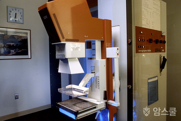
유방조영술의 한계
유방조영촬영검사는 현재 우리가 가지고 있는 유방암 검사 중 최고의 검사입니다. 하지만 유방조영술은 한계가 있습니다. 예를 들어, 여성의 유방암 여부를 100% 정확하게 보여주지 못합니다.
거짓 음성 유방조영술은 유방암이 존재해도 정상으로 보입니다.
거짓 양성 유방조영술은 유방에 암이 없는데도 비정상적으로 보입니다.
거짓 음성 화상
거짓 음성 유방조영술은 유방암이 존재해도 정상으로 보입니다. 전반적으로, 유방조영촬영검사는 5개의 유방암 중 1개를 발견하지 못합니다.
치밀 유방 여성은 거짓 음성 결과가 나올 확률이 높습니다.
거짓 음성 유방조영술은 여성들에게 사실 유방암에 걸리지 않았다고 생각하는 잘못된 안정감을 줄 수 있습니다.
거짓 양성 화상
거짓 양성 유방조영술은 암이 실제로 존재하지 않더라도 비정상적으로 보입니다. 비정상적인 유방조영술은 종종 그 변화가 암인지 알아보기 위해 추가 검사(진단 유방조영술, 초음파 검사, 때로는 MRI나 심지어 유방 생체검사)가 필요합니다.
거짓 양성 화상은 나이가 어리고, 유방이 치밀하며, 유방 생체검사를 받았거나, 가족 내에서 유방암을 앓거나, 에스트로겐을 복용하고 있는 여성들에게 더 흔합니다.
10년 동안 매년 유방조영술을 받는 여성의 약 절반은 언젠가는 거짓 양성 판정을 받게 될 것입니다.
첫 번째 유방조영술의 경우 거짓 양성 소견의 확률이 가장 높습니다. 비교 가능한 과거 유방조영술을 받은 여성은 거짓 양성 소견의 확률을 약 50%까지 낮춥니다.
거짓 양성 유방조영술은 불안감을 유발할 수 있습니다. 그들은 또한 암이 없다는 것을 확실히 하기 위한 추가 검사로 이어질 수 있습니다. 그것은 시간과 비용, 그리고 심지어 육체적인 불편함까지도 필요로 합니다.
유방조영술은 모든 여성에게 도움이 되지 않을 수 있습니다.
유방 촬영 검진의 가치는 여성의 전반적인 건강에 따라 달라집니다. 유방암을 조기에 발견하는 것은 그녀가 심각한 심장 질환이나 심각한 신장, 간 또는 폐 질환과 같은 다른 심각하거나 생명을 위협하는 건강 문제를 야기한다면 더 오래 사는 데 도움이 되지 않을 수 있습니다. 미국 암 협회의 유방암 검진 지침은 심각한 건강 문제나 기대 수명이 짧은 여성들이 유방조영술을 계속 받아야 하는지에 대해 의사와 상의해야 한다고 강조합니다. 우리의 지침은 또한 나이만으로 정기적인 유방조영술을 중단하는 이유가 되어서는 안 된다고 강조합니다.
유방조영술은 종종 너무 작아서 느낄 수 없는 유방암을 발견할 수 있지만, 작은 종양을 치료한다고 해서 항상 완치가 되는 것은 아니라는 것을 아는 것이 중요합니다. 빠르게 성장하거나 공격적인 암이 이미 퍼졌을지도 모릅니다.
과잉 진단 및 과잉 치료
유방조영촬영검사는 종종 치료가 필요한 침습성 유방암과 유방상피내암(DCIS)을 현장에서 발견할 수 있습니다. 그러나 유방조영술에서 발견된 일부 침습성 암과 DCIS는 결코 성장하거나 확산되지 않을 가능성이 있습니다.(절대 문제를 일으키지 않는 암을 찾아 치료하는 것을 과잉진단이라고 합니다.) 이 암들은 생명을 위협하지 않으며, 만약 여성이 유방조영술을 받지 않았다면 발견되거나 치료되지 않았을 것입니다. 문제는 의사들이 이러한 암이 자라고 퍼질 암과 구별할 수 없다는 것입니다.
과잉진단은 일부 여성들에게 별로 필요하지 않은 치료를 받게 합니다. 왜냐하면 암은 어떤 문제도 일으키지 않았을 것이기 때문입니다. 의사들은 어떤 암이 생명을 위협하고 어떤 암이 결코 문제를 일으키지 않는지 항상 알 수는 없습니다. 이 때문에 모든 사례에 대해 치료를 합니다. 이것은 몇몇 여성들에게 암 치료의 부작용에 노출시킵니다. 비록 그것이 정말로 필요하지 않더라도 말이죠.
하지만, 과잉 진단은 그리 자주 일어나지 않을 것으로 생각됩니다. 유방암 중 유방조영술에 의해 과잉 진단될 수 있는 비율에 대한 광범위한 추정치가 있지만, 가장 신뢰할 수 있는 추정치는 1%에서 10%까지 다양합니다.
방사선 노출
유방조영술은 X선 검사이기 때문에 유방을 방사선에 노출시킨다. 각 유방조영술의 방사선량은 낮지만 시간이 지남에 따라 증가될 수 있습니다. 자세한 내용은 유방 촬영 기본사항을 참조하시기 바랍니다.
최신 의료 리뷰 : 2019년 10월 3일
Limitations of Mammograms
Last Medical Review: October 3, 2019 Last Revised: October 3, 2019
Mammograms are the best breast cancer screening tests we have at this time. But mammograms have their limits. For example, they aren’t 100% accurate in showing if a woman has breast cancer:
A false-negative mammogram looks normal even though breast cancer is present.
A false-positive mammogram looks abnormal even though there’s no cancer in the breast.
False-negative results
A false-negative mammogram looks normal even though breast cancer is present. Overall, screening mammograms do not find about 1 in 5 breast cancers.
Women with dense breasts are more likely to get false-negative results.
False-negative mammograms can give women a false sense of security, thinking that they don’t have breast cancer when in fact they do.
False-positive results
A false-positive mammogram looks abnormal even though no cancer is actually present. Abnormal mammograms often require extra testing (diagnostic mammograms, ultrasound, and sometimes MRI or even a breast biopsy) to find out if the change is cancer.
False-positive results are more common in women who are younger, have dense breasts, have had breast biopsies, have breast cancer in the family, or are taking estrogen.
About half of the women getting annual mammograms over a 10-year period will have a false-positive finding at some point.
The odds of a false-positive finding are highest for the first mammogram. Women who have past mammograms available for comparison reduce their odds of a false-positive finding by about 50%.
False-positive mammograms can cause anxiety. They can also lead to extra tests to be sure cancer isn’t there, which cost time and money and maybe even physical discomfort.
Mammograms might not be helpful for all women
The value of a screening mammogram depends on a woman’s overall health. Finding breast cancer early may not help her live longer if she has other serious or life-threatening health problems, such as serious heart disease, or severe kidney, liver, or lung disease. The American Cancer Society breast cancer screening guidelines emphasize that women with serious health problems or short life expectancies should discuss with their doctors whether they should continue having mammograms. Our guidelines also stress that age alone should not be the reason to stop having regular mammograms.
It’s important to know that even though mammograms can often find breast cancers that are too small to be felt, treating a small tumor does not always mean it can be cured. A fast-growing or aggressive cancer might have already spread.
Overdiagnosis and overtreatment
Screening mammograms can often find invasive breast cancer and ductal carcinoma in situ (DCIS, cancer cells in the lining of breast ducts) that need to be treated. But it’s possible that some of the invasive cancers and DCIS found on mammograms would never grow or spread. (Finding and treating cancers that would never cause problems is called overdiagnosis.) These cancers are not life-threatening, and never would have been found or treated if the woman had not gotten a mammogram. The problem is that doctors can’t tell these cancers from those that will grow and spread.
Overdiagnosis leads to some women getting treatment that’s not really needed (overtreatment), because the cancer never would have caused any problems. Doctors can’t always tell which cancers will be life-threatening and which won’t ever cause problems. Because of this, all cases are treated. This exposes some women to the side effects of cancer treatment, even though it’s really not needed.
Still, overdiagnosis is not thought to happen very often. There’s a wide range of estimates of the percentage of breast cancers that might be overdiagnosed by mammography, but the most credible estimates range from 1% to 10%.
Radiation exposure
Because mammograms are x-ray tests, they expose the breasts to radiation. The amount of radiation from each mammogram is low, but it can still add up over time. For more on this, see Mammogram Basics.
이 글은 2020년 6월 작성 시점의 정보입니다. 치료 기준·급여 정보는 바뀌었을 수 있으니, 진료와 치료 결정은 반드시 담당 의료진과 상의하세요.