인공 지능으로 수술 중 뇌종양 진단의 신속화 가능.
2020.03.03 · 전영조 전문위원
인공 지능으로 수술 중 뇌종양 진단의 신속화 가능
2020년 2월 12일 : 국립암연구소 제공
뇌종양이 있는 환자의 경우, 치료의 첫 번째 단계는 종양을 최대한 많이 제거하는 수술이다. 수술 중에 획득하고 분석한 종양 샘플은 종양을 정밀하게 진단하고 종양과 건강한 뇌 조직 사이의 경계를 밝혀내는 데 도움을 줄 수 있다.
그러나 이러한 수술 중 병리학 분석에는 시간이 걸린다. 즉, 외과의사와 환자가 결과를 기다리는 동안 병리학자가 샘플을 처리하고, 염색하고, 분석해야 한다. 이제 새로운 연구는 첨단 영상 기술과 인공지능(AI)을 결합한 과정을 통하여 수술 중 3분 이내에 뇌종양을 정확하게 진단할 수 있다는 것을 보여준다. 이 접근법은 또한 종양 조직과 건강한 조직을 정확하게 구별할 수 있었다.
이 연구결과는 1월 6일 ‘네이처 메디신’ 지에 발표되었다.
이번 연구를 주도한 뉴욕대학 랭곤 헬스의 대니얼 오링거 박사는 "이 기술은 특히 새로 발견된 종양 환자와 2차나 3차 수술을 받고 있는 재발 환자들에게 고무적인 기술"이라고 말했다.
연구팀은 이 연구가 "수술 중 즉석으로 수술에 필요한 조직 진단에 비할 데 없이 접근할 수 있는 기회를 제공하는 한편, "종양에 인접한 정상 조직을 제거할 위험을 줄인다."고 썼다.
연구에 참여하지 않은 NIH의 외과 신경학 부의 신경외과 의사인 카림 자글루 박사는 이번 연구 결과에 고무되어 있다고 말했다. "이 기술은 수술이 얼마나 공격적이거나 보수적일 필요가 있는지 알려주는 데 도움이 될 것"이라고 자글루 박사는 말했다.
화상 및 AI 기술의 적용
이 연구에서 미시건 대학의 수석 신경외과 전문의인 오링거 박사와 토드 홀론 박사가 이끄는 연구팀은 stimulated Raman histology (SRH)라는 영상기술을 AI의 예측력과 결합시켜 현재의 수술 중 행하는 병리학 관행을 개선할 수 있는지 실험해 보고자 했다.
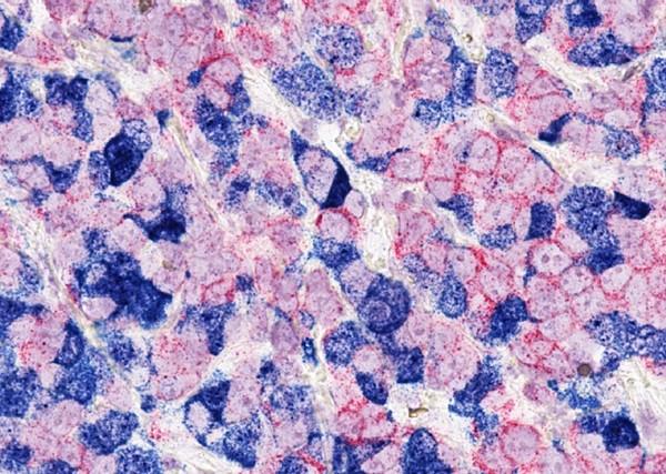
특수화된 형태의 현미경 관찰법인 SRH는 수술실에서 직접 신선한 조직 샘플을 시각화하는 데 사용될 수 있으며, 심지어 병리학자들이 세포 구조를 분석하기 위해 냉동 조직 샘플에 이용하는 것과 같은 종류의 "착색"을 생성하기도 한다. 미시간 대학에서 외과 팀은 이미 일부 뇌종양과 두경부 암 시술에 SRH 시스템을 사용하고 있다.
인공지능은 일반적으로 인간의 지능과 관련된 업무를 수행하기 위해 강력한 컴퓨터를 사용하는 것을 포함한다. 심층학습으로 알려진 AI의 한 종류는 복잡한 수학적 알고리즘을 사용하는데, 때로는 중첩신경망이라고도 한다. 이때 그것이 "훈련된" 데이터로부터 형상을 추출하기 위해서다.
이 훈련은 알고리즘이 패턴을 인식하고 영상 분석 등의 작업을 수행할 수 있도록 한다. 예를 들어 의학에서는 그러한 알고리즘이 유방조영술을 평가하거나, 자궁경부의 전 암 조직을 검출하거나, 암 분자를 보다 정확하게 검출할 수 있는지 알아보기 위해 연구되고 있다.
SRH 이미저(영상 기기)의 힘을 AI와 결합하기 위해, 연구자들은 뇌종양 조직의 SRH 생산 영상에 대한 알고리즘을 훈련하는 것으로 시작했다. 이 훈련을 위해 그들은 415명의 환자의 250만 개 이상의 종양 조직 이미지(영상)를 사용했다. 이 이미지는 건강한 회색 또는 백색 물질을 포함한 세 가지의 비 종양으로 분류된 것과 미국에서 모든 뇌종양 진단 중 90% 이상을 차지하는 10 가지의 가장 흔한 뇌종양 유형을 포함했다.
홀론 박사는 "알고리즘을 훈련시키기 위한 이상적인 크기와 해상도를 결정하는 것이 초기 과제였다"고 말했다. 이러한 이상적인 매개변수가 결정되면, 알고리즘은 조직 표본을 최종 종양, 비 종양 조직 또는 비 진단(AI로 분석할 수 없다는 뜻) 조직으로 분류하는 법을 배웠다.
임상 시험에서 테스트
뇌종양 진단을 위한 SRH‒AI 기술의 임상적 가치를 탐구하기 위해 연구원들은 280명에 가까운 환자를 임상시험에 등록시켰는데, 이들 모두는 외과 의사들이 보통 수집되는 것 이상의 추가적인 종양 조직을 추가로 수집할 수 있도록 허용하는데 동의했다.
조직표본을 2개로 나누어 수술실의 신기술(알고리즘에 의해 분류된 신기술(SRH 영상)과 종래의 실험실 병리검사(병리학자가 처리, 염색, 분석)를 이용하여 분석하여 신기술이 기존 기술만큼 정확한지 확인하였다.
그것이 사실임이 판명되었다. 이 알고리즘은 뇌종양을 94.6%의 정확도로 진단한 반면, 종래의 병리학자 기반 분석은 93.9%의 전체 정확도를 보였다.
조직 표본이 알고리즘에 의해 잘못 분류된 경우, 병리학자는 정확한 진단을 내렸다. 그리고 병리학자들이 샘플을 잘못 분류한 경우, 알고리즘은 정확한 진단을 내렸다.
연구원들은 AI 기술과 병리학자들이 상호 점검할 수 있는 능력은 병리학자들이 어려운 사례를 해석하고 가능한 최고의 진단 정확도를 보장하기 위해 AI 기술과 함께 일할 필요성을 강조한다는 점에 주목했다.
수술의 정확도 향상
종양 절제의 정도는 수술 후 MRI 스캔을 통해서도 알 수 있으며, 수술 후 절제가 얼마나 완전했는지 알 수 있다. 수술 중에 가능한 한 많은 종양을 제거하면 환자의 수명을 향상시킬 수 있지만 수술 중에 너무 많은 건강한 뇌 조직을 제거하면 운동 기능 저하, 기억 상실 또는 시력 장애와 같이 환자에게 심각하고 해로운 결과를 초래할 수 있다.
이를 해결하기 위해 연구진은 환자가 아직 수술이 진행되는 동안 건강한 뇌 조직과 종양 조직을 구별할 수 있는 신기술의 능력도 조사하였다.
종양세포는 때때로 건강한 조직에 침투할 수 있기 때문에 수술 중 육안으로 종양과 건강한 조직의 경계를 식별하기 어려울 수 있다. AI 기술은 각 표본 이미지를 더 작은 ‘패치’로 분해해 외과 의사들이 종양이나 건강한 조직을 포함한 부위를 빠르고 명확하게 확인할 수 있도록 했다.
SRH‒AI 기술을 사용하여, 오링거 박사는 "수술 중에 보이지 않을 종양[세포]을 시각화할 수 있다"고 말했다.
그리고 "환자의 예후는 절제 정도에 따라 달라지기 때문에, 뇌와 종양의 경계선에 대한 더 나은 정보를 가지고 있는 자글루 박사는 "환자에게 더 나은 치료 결과를 가져올 수 있고 건강한 조직이 보존되어 있기 때문에 수술과 관련된 합병증이 줄어들 수 있다"고 말했다.
임상 시험에서 테스트
뇌종양 진단을 위한 SRH‒AI 기술의 임상적 가치를 탐구하기 위해 연구원들은 280명에 가까운 환자를 임상시험에 등록시켰는데, 이들 모두는 외과의사들이 보통 수집되는 것 이상의 추가적인 종양 조직을 추가로 수집할 수 있도록 허용하는데 동의했다.
조직표본을 2개로 나누어 수술실의 신기술(알고리즘에 의해 분류된 신기술(SRH 영상)과 종래의 실험실 병리(병리학자가 처리, 염색, 분석)를 이용하여 분석하여 신기술이 기존 기술만큼 정확한지 확인하였다.
그것이 사실임이 판명되었다. 이 알고리즘은 뇌종양을 정확하게 진단한 반면, 종래의 병리학자 기반 분석은 93.9%의 전체 정확도를 보였다.
조직 표본이 알고리즘에 의해 잘못 분류된 경우, 병리학자는 정확한 진단을 내렸다. 그리고 병리학자들이 샘플을 잘못 분류한 경우, 알고리즘은 정확한 진단을 내렸다.
연구원들은 AI 기술과 병리학자들이 상호 점검할 수 있는 능력은 병리학자들이 어려운 사례를 해석하고 가능한 최고의 진단 정확도를 보장하기 위해 AI 기술과 함께 일할 필요성을 강조한다는 점에 주목했다.
수술 정확도 향상
종양 제거의 정도는 수술 후 MRI 스캔을 통해서도 알 수 있으며, 수술 후 제거가 얼마나 완료되었는지를 알 수 있다. 수술 중에 가능한 한 많은 종양을 제거하면 환자의 수명이 단축되지만 수술 중에 너무 많은 건강한 뇌 조직을 제거하면 운동 기능 저하, 기억 상실 또는 시력 장애와 같이 환자에게 심각하고 해로운 결과를 초래할 수 있다.
이를 해결하기 위해 연구진은 환자가 아직 수술 중인 동안 건강한 뇌 조직과 종양 조직을 구별할 수 있는 신기술의 능력도 조사했다.
종양세포는 때때로 건강한 조직에 침투할 수 있기 때문에 수술 중 육안으로 종양과 건강한 조직의 경계를 식별하기 어려울 수 있다. AI 기술은 각 표본 이미지를 더 작은 ‘패치’로 분해해 외과의사들이 종양이나 건강한 조직을 포함한 부위를 빠르고 명확하게 확인할 수 있도록 했다.
SRH‒AI 기술을 사용하여, 오링거 박사는 "수술 중에 보이지 않을 종양[세포]을 시각화할 수 있다"고 말했다.
그리고 "환자의 예후는 절제 정도에 따라 달라지기 때문에, 뇌척수 마진에 대한 더 나은 정보를 가지고 있는 자글루 박사는 "환자에게 더 나은 치료 결과를 가져올 수 있고 건강한 조직이 보존되어 있기 때문에 수술과 관련된 합병증이 줄어들 수 있다"고 말했다.
뇌종양 관련 AI(인공지능)의 미래
새로운 기술이 다른 병원과 기관으로 확장되기 전에, "더 많은 환자들을 대상으로 한 강력한 테스트와 희귀한 뇌종양을 포함하는 기술의 확대가 매우 필요하다"고 자글루 박사는 말했다.
SRH 이미저(영상 기기)는 오늘날 미국 전역의 몇몇 주요 암 센터에서 사용되고 있다. 오리너 박사는 "AI와 SRH 화상기술은 모두 신흥 기술이기 때문에 이를 진료에 통합하는 데 어려움이 있을 것"이라고 설명했다.
그렇더라도, 오링거 박사는 SRH‒AI 기술의 사용이 향후에는 병리학적 자원이 제한된 센터와 여러 가지 다른 종류의 암에 잠재적으로 사용할 수 있도록 확장될 것이라고 희망하고 있다.
Artificial Intelligence Expedites Brain Tumor Diagnosis during Surgery
February 12, 2020, by NCI Staff
For patients with a brain tumor, the first step in treatment is often surgery to remove as much of the mass as possible. A tumor sample obtained and analyzed during surgery can help to precisely diagnose the tumor and define the margins between tumor and healthy brain tissue.
However, such intraoperative pathology analysis takes time—the sample must be processed, stained, and analyzed by a pathologist while the surgeon and patient wait for the results. Now, a new study shows that a process combining an advanced imaging technology and artificial intelligence (AI) can accurately diagnose brain tumors in fewer than 3 minutes during surgery. The approach was also able to accurately distinguish tumor tissue from healthy tissue.
The findings were published January 6 in Nature Medicine.
“This technology is especially encouraging for patients with newly detected tumors and patients with [recurrent tumors] who are undergoing second or third surgeries,” said Daniel Orringer, M.D., of NYU Langone Health, who helped lead the study.
This study, the research team wrote, opens the door to “provid[ing] unparalleled access to intraoperative tissue diagnosis at the bedside during surgery” while “reducing the risk of removing … normal tissue adjacent to a [tumor].”
Kareem Zaghloul, M.D., Ph.D., a neurosurgeon in NIH’s Surgical Neurology Branch who was not involved in the research, said he is encouraged by the study’s results. “This technology could help inform how aggressive or conservative surgery needs to be,” Dr. Zaghloul said.
Applying Imaging and AI Technology
In the study, a research team led by Dr. Orringer and Todd Hollon, M.D., chief neurosurgery resident at the University of Michigan, wanted to test whether they could combine an imaging technology called stimulated Raman histology (SRH) with the predictive power of AI to improve current intraoperative pathology practice.
SRH, a specialized form of microscopy, can be used to visualize fresh tissue samples directly in the operating room, even producing the same sort of “staining” that pathologists apply to frozen tissue samples to analyze cellular structure. At the University of Michigan, surgical teams are already using an SRH system for some brain tumor and head and neck cancer procedures.
AI involves using powerful computers to perform tasks that are typically associated with human intelligence. A type of AI known as deep learning uses complex mathematical algorithms, sometimes called convolutional neural networks, to extract features from data that it is then “trained” on.
This training allows the algorithm to recognize patterns and perform tasks such as analyzing images. In medicine, for example, such algorithms are being studied to see if they can help assess mammograms, detect precancerous tissue in the cervix, or detect cancerous moles more accurately.
To combine the power of the SRH imager with AI, the researchers began by training an algorithm on SRH-produced images of brain tumor tissue. For the training, they used more than 2.5 million tumor tissue images from 415 patients. The images covered three nontumor-tissue classifications, including healthy grey or white matter, and the 10 most common brain tumor types, which account for more than 90% of all brain tumor diagnoses in the United States.
“A major initial challenge was determining the ideal size and resolution of images to train the algorithm,” Dr. Hollon said. Once these ideal parameters were determined, the algorithm learned to classify tissue samples as definitive tumor, nontumor tissue, or nondiagnostic (meaning they couldn’t be analyzed by AI).
Testing in a Clinical Trial
To explore the clinical value of the SRH‒AI technology for diagnosing brain tumors, the researchers enrolled nearly 280 patients in a clinical trial, all of whom agreed to allow surgeons to collect additional tumor tissue beyond what would usually be collected but in a way that would not pose additional risks.
Tissue specimens were divided into two and analyzed using the new technology (SRH images classified by the algorithm) in the operating room and conventional laboratory pathology (tissues processed, stained, and analyzed by a pathologist) to see if the new technology was as accurate as the conventional technology.
That proved to be the case. The algorithm correctly diagnosed brain tumors 94.6% of the time, while conventional pathologist-based analysis had an overall accuracy rate of 93.9%.
In the instances where tissue specimens were incorrectly classified by the algorithm, a pathologist had made the correct diagnosis. And in the instances where pathologists incorrectly classified a sample, the algorithm had made the correct diagnosis.
The researchers noted that the ability of the AI technology and pathologists to cross-check each other highlights the need for pathologists to work alongside the AI technology to interpret challenging cases and ensure the highest diagnostic accuracy possible.
Improving Surgical Accuracy
The extent of tumor removal can be determined during surgery as well as with a post-operative MRI scan that shows how complete the removal was. While removing as much tumor as possible during surgery can improve how long patients live, removing too much healthy brain tissue during surgery can have serious and harmful consequences for a patient, such as impaired motor function, memory loss, or vision problems.
To address this, the researchers also looked at the ability of the new technology to distinguish tumor tissue from healthy brain tissue while the patient is still in surgery.
Because tumor cells can sometimes infiltrate healthy tissues, it can be difficult to identify the border between tumor and healthy tissue with the naked eye during surgery. By breaking up each specimen image into smaller “patches,” the AI technology allows surgeons to quickly and clearly identify areas containing tumor or healthy tissue.
Using SRH‒AI technology, Dr. Orringer said, “we can … visualize tumor [cells] that would otherwise be invisible during surgery.”
And since “a patient’s prognosis is dependent on the extent of resection,” said Dr. Zaghloul, having better information on the brain‒tumor margin “could result in better treatment outcomes for patients and fewer surgery-related complications due to healthy tissue being preserved.”
The Future of AI in Brain Tumors
Before the new technology can be expanded to other centers and institutions, “robust testing with more patients and expanding the technology to include rare brain tumors are greatly needed,” Dr. Zaghloul said.
The SRH imager is being used at several major cancer centers across the United States today. Both AI and SRH imaging are emerging technologies, so there will be challenges to integrating them into care, Dr. Orringer explained, such as financial or regulatory issues as well as clinician training.
Even so, Dr. Orringer is hopeful that use of the SRH‒AI technology will expand in the future, including at centers with limited pathology resources and for potential use in a number of different cancer types.
이 글은 2020년 3월 작성 시점의 정보입니다. 치료 기준·급여 정보는 바뀌었을 수 있으니, 진료와 치료 결정은 반드시 담당 의료진과 상의하세요.