췌장 신경내분비종양 치료 종합정보
2021.01.20 · 전영조 전문위원

1. 췌장 신경내분비종양(섬세포 종양)에 대한 일반 정보
췌장 신경 내분비 종양은 췌장의 호르몬 생성 세포에서 형성된다. 췌장은 옆으로 누운 얇은 배처럼 생긴 약 6인치 길이의 분비선이다. 췌장의 넓은 끝은 머리, 중간 부분은 몸체, 그리고 좁은 끝은 꼬리라고 부른다. 췌장은 위의 뒤와 척추 앞에 놓여 있다.
췌장에는 두 가지 종류의 세포가 있다
• 내분비 췌장 세포는 혈당을 조절하기 위한 인슐린과 같은 여러 종류의 호르몬(인체의 특정 세포나 장기의 작용을 조절하는 화학 물질)을 만든다. 그들은 췌장 전체에 걸쳐 많은 작은 집단으로 함께 모여있다. 내분비 췌장 세포는 섬 세포 혹은 랑게르한스 섬으로 불린다. 섬 세포에서 형성되는 종양을 섬 세포 종양, 췌장 내분비종양 또는 췌장 신경내분비종양이라고 부른다.
• 외분비 췌장 세포는 소장으로 방출되는 효소를 만들어 신체가 음식을 소화하는 것을 돕는다. 대부분의 췌장은 관의 끝에 작은 주머니가 있는 도관으로 이루어져있는데, 이 도관은 외분비 세포로 내벽이 구성되어 있다.
췌장 신경 내분비 종양(NETs)은 양성(암이 아닌) 또는 악성(암)일 수 있다. 췌장 NET가 악성일 때, 그것들은 췌장 내분비암 또는 섬 세포 암이라고 부른다.
췌장 신경내분비 종양(NET)은 췌장 외분비 종양보다 훨씬 덜 흔하고 더 나은 예후를 가지고 있다. 췌장 NET는 징후나 증상을 유발하거나 혹은 유발하지 않을 수 있다.
췌장 NET는 기능성 종양(호르몬을 생성)이거나 비 기능성(호르몬을 생성하지 못함) 종양일 수 있다.
• 기능성 종양은 가스트린, 인슐린, 글루카곤과 같은 호르몬을 추가로 만들어 증상과 징후를 유발한다.
• 비 기능성 종양은 여분의 호르몬을 생성하지 않는다. 증상과 징후는 종양이 퍼지고 자라면서 생기는 것이다. 대부분의 비기능성 종양은 악성종양이다.
대부분의 췌장 NET은 기능성 종양이다. 기능성 췌장 NET에는 다양한 종류가 있다. 췌장 NET는 가스트린, 인슐린, 글루카곤과 같은 다른 종류의 호르몬을 만든다. 기능 췌장 NET에는 다음이 포함된다.
• 가스트린종 : 가스트린을 만드는 세포에서 형성되는 종양. 가스트린은 위가 음식 소화를 돕는 산을 방출하도록 하는 호르몬이다. 가스트린과 위산은 둘 다 가스트린종에 의해 증가된다. 위산과다와 위궤양, 설사가 가스트린을 만드는 종양에 의해 야기될 때, 이것은 졸링거 엘리슨 증후군이라고 부른다. 위염은 보통 췌장의 머리에서 형성되며 때로는 소장에서 형성되기도 한다. 대부분의 가스트린종은 악성종이다.
• 인슐린종 : 인슐린을 만드는 세포에서 형성되는 종양. 인슐린은 혈중 포도당의 양을 조절하는 호르몬이다. 그것은 포도당을 세포로 이동시키고, 거기서 그것은 신체가 에너지를 위해 사용할 수 있다. 인슐린 종양은 보통 잘 퍼지지 않는 천천히 자라는 종양이다. 인슐린종은 췌장의 머리, 몸 또는 꼬리 부분에 형성된다. 인슐린종은 대개 양성이다.
• 글루카곤종 : 글루카곤을 만드는 세포에서 형성되는 종양. 글루카곤은 혈중 포도당의 양을 증가시키는 호르몬이다. 간은 글리코겐을 분해한다. 너무 많은 글루카곤은 고혈당증을 유발한다. 글루카곤종은 보통 췌장의 꼬리에서 형성된다. 대부분의 글루카곤종은 악성이다.
• 기타 종양 유형 : 호르몬을 만드는 다른 희귀한 유형의 기능성 췌장 NET들이 있는데, 여기에는 당분, 소금, 그리고 체내의 물의 균형을 조절하는 호르몬이 포함된다. 이러한 종양에는 다음이 포함된다. :
- 혈관 활성 장 펩타이드를 만드는 비포마스 비포마는 또한 베르너-모리슨 증후군이라고도 불릴 수 있다.
- 소마토스타틴종은 소마토스타틴을 만든다.
※ VIPoma비포마 : 내분비종양으로서, 일반적으로 비장(脾臟)으로부터 기원하며, 혈관작용성 장관 폴리펩타이드(vasoactive intestinal polypeptide)를 생산한다. 이 물질은 강한 심혈관 및 전해질 변화를 일으켜 혈관확장성 저혈압, 수성설사, 저 칼슘혈증 및 탈수를 가져온다고 믿어진다.
이러한 다른 종류의 종양들은 같은 방식으로 치료되기 때문에 함께 분류된다. 특정 증후군이 있으면 췌장 NET의 위험을 증가시킬 수 있다. 당신이 질병에 걸릴 위험을 증가시키는 모든 것을 위험 인자(요소)라고 부른다. 위험인자가 있다는 것이 암에 걸린다는 것을 의미하지 않고 위험인자가 없다고 암에 걸리지 않는다는 것을 의미하지 않는다.
위험하다고 생각되면 의사와 상담하라. 다발성 내분비 신생물 1형 증후군은 췌장 NET의 위험인자이다. 췌장 NET의 유형별로 증상과 징후가 다르다. 징후나 증상은 종양의 성장이나 그 종양이 만드는 호르몬이나 다른 질환에 의해 야기될 수 있다. 일부 종양은 징후나 증상을 일으키지 않을 수 있다. 이러한 문제가 있으면 의사에게 문의하시오.
비 기능성 췌장 신경내분비 종양의 징후 및 증상
비기능성 췌장 NETSMS 징후나 증상을 일으키지 않고 오랫동안 증식할 수 있다. 다음과 같은 증상이나 징후를 일으키기 전에 커지거나 신체의 다른 부분으로 퍼질 수 있다.
• 설사
• 소화불량
• 복부에 혹
• 복부나 등에 통증
• 피부나 눈의 흰자위의 황달
췌장 기능성 NET(신경내분비 종양)의 징후 및 증상
기능성 췌장 NET의 징후와 증상은 만들어지는 호르몬의 종류에 따라 달라진다. 가스트린이 너무 많으면 다음을 유발할 수 있다.
• 계속 재발하는 위궤양
• 등 쪽으로 퍼질 수 있는 복부의 통증. 통증은 생겼다 사라질 수도 있고 제산제를 사용하면 통증이 사라질 수 있다.
• 위 내용물이 식도로 재 역류(위상 역류)
• 설사
인슐린 과다 분비는 다음을 유발할 수 있다
• 저혈당. 이것은 시야의 혼탁, 두통, 그리고 현기증, 피곤, 쇠약, 떨림, 긴장감, 짜증, 땀, 혼란 또는 배고픔을 유발할 수 있다.
• 빠른 심장 박동
글루카곤이 너무 많으면 다음이 발생할 수 있다
• 피부발진. 특히 얼굴과 위 또는 다리에 발진
• 고혈당. 이것은 두통, 잦은 배뇨, 피부와 구강건조, 또는 허기, 갈증, 피로 혹은 쇠약을 유발 할 수 있다.
• 혈전. 폐에 혈전이 생기면 호흡 곤란과 기침, 가슴 통증 등이 생길 수 있다. 팔이나 다리에 혈전이 생기면 통증과 부종, 열기 또는 팔이나 다리의 붉은색이 나타날 수 있다.
• 설사
• 이유를 알 수 없는 체중 감소
• 입의 모서리에 혀나 상처
혈관 활성 장 펩타이드(VIP)가 너무 많으면 다음이 발생할 수 있다
• 다량의 물 설사
• 탈수. 이것은 갈증을 느끼거나, 소변을 적게 만들거나, 피부와 입이 건조하거나, 두통, 현기증, 또는 피곤함을 느끼게 할 수 있다.
• 혈액 내 칼륨 수치의 저하. 이것은 근육 약화, 통증 또는 경련, 저림과 얼얼함, 잦은 배뇨, 빠른 심장박동, 혼돈이나 갈증을 유발할 수 있다.
• 복부 경련 또는 통증
• 알 수 없는 이유로 체중 감소
소마토스타틴이 너무 많으면 다음을 유발할 수 있다
• 고혈당. 이것은 두통, 잦은 배뇨, 건조한 피부와 입, 또는 배고프거나 목마르고 피곤하거나 허약함을 느끼게 할 수 있다.
• 설사
• 지방변증(물에 뜨는 냄새가 매우 고약한 변)
• 담석
• 피부와 눈의 흰자위 황달
• 이유를 알 수 없는 체중감소
췌장 NET는 또한 너무 많은 부신피질 호르몬을 만들어 쿠싱 증후군을 일으킬 수 있다. 쿠싱 증후군의 징후와 증상은 다음과 같다.
• 두통
• 일부 시력 상실
• 얼굴과 목, 몸통의 체중 증가, 팔과 다리가 가늘어짐
• 목 뒤쪽의 지방 덩어리
• 가슴이나 복부에 보라색 또는 분홍색 스트레치 자국이 생길을 수 있는 얇은 피부
• 쉽게 드는 멍
• 얼굴, 등의 윗부분 또는 팔에 미세한 털
• 쉽게 부러지는 뼈
• 치유가 느린 염증이나 상처
• 불안, 짜증, 우울증
너무 많은 부신피질 자극 호르몬과 Cushing 증후군을 만드는 췌장 NET의 치료는 이 요약에서 논의되지 않았다. 실험실 검사와 영상 검사는 췌장 NET를 감지(발견)하고 진단하는 데 사용된다. 다음 검사 및 시술을 할 수 있다.
• 신체검사 및 이력 : 혹 등 질환의 징후를 확인하는 등 일반적인 건강의 징후를 확인하기 위한 신체검사. 환자의 건강 습관과 과거의 질병과 치료의 기록도 반영한다.
• 혈액 화학 연구 : 혈액을 검사하여 혈당과 같은 특정 물질의 양을 신체 내 장기 및 조직에 의해 혈액으로 방출되는 양을 측정하는 시술. 물질의 양이 비정상적이면 질병의 징후일 수 있다.
• 크로모그래닌 A 검사 : 혈액 내 크로모그래닌 A의 양을 측정하기 위해 혈액 샘플을 검사하는 검사. 가스트린, 인슐린, 글루카곤과 같은 크로모그래닌의 양과 정상 호르몬의 양이 정상보다 많으면 비 기능성 췌장 NET의 징후가 될 수 있다.
• 복부 CT 스캔(CAT 스캔) : 여러 각도에서 촬영한 복부의 세부 사진을 연속으로 만드는 시술. 그 사진들은 엑스레이 기계에 연결된 컴퓨터로 만들어진다. 조영제인 염료는 정맥에 주입되거나 기관이나 조직이 더 명확하게 나타나도록 복용할 수 있다. 이 절차는 컴퓨터 단층 촬영, 또는 컴퓨터화 된 축 단층 촬영이라고도 한다.
• MRI(자기 공명 영상) : 자석, 전파, 컴퓨터를 사용하여 신체 내부의 여러 부위를 세밀하게 촬영하는 시술. 이 시술을 NMRI(핵자기 공명 영상)라고도 한다.
• 소마토스타틴 수용체 신티그램 : 소형 췌장 NET를 찾는 데 사용할 수 있는 방사성핵종 스캔 유형. 소량의 방사성 옥트로타이드(종양에 부착되는 호르몬)가 정맥에 주입돼 혈액을 통해 이동한다. 방사성 옥트레오티드는 종양에 부착되고 방사능을 감지하는 특수 카메라는 종양이 몸 안에 어디에 있는지를 보여주기 위해 사용된다. 이 시술을 옥트레오티드 스캔 및 SRS라고도 부른다.
• 내시경 초음파(EUS) : 내시경을 신체에 삽입하는 시술로서, 보통 입이나 직장을 통해 삽입한다. 내시경은 빛과 렌즈가 부착된 얇고 튜브 같은 기구이다. 내시경 끝의 탐침은 내부 조직이나 장기에서 고에너지 음파(초음파)를 튕겨내고 메아리를 만드는 데 사용된다. 메아리는 초음파영상이라는 신체 조직의 그림을 형성한다. 이 시술은 내시경초음파 검사라고도 부른다.
• 내시경 역행담췌관조영(ERCP) : 담낭에서 소장으로 담즙을 운반하는 관(튜브)을 X선 촬영하는 시술. 때때로 췌장암은 이러한 관을 좁히고 담즙의 흐름을 막거나 느리게 하여 황달을 유발한다. 내시경은 입, 식도, 위를 거쳐 소장의 첫 부분으로 전달된다. 내시경은 빛과 렌즈를 가진 얇고 튜브 같은 기구이다. 그런 다음 카테터(작은 튜브)가 내시경을 통해 췌장관으로 삽입된다. 카테터를 통해 도관으로 염료가 주입되고 X선이 촬영된다. 종양으로 인해 도관이 막힌 경우 미세한 튜브를 도관에 삽입하여 차단을 해제할 수 있다. 이 튜브(또는 스텐트)는 췌관을 열린 상태로 유지하기 위해 그대로 둘 수 있다. 조직 샘플은 현미경으로 관찰하여 암의 징후를 확인할 수도 있다.
• 혈관조영법 : 혈관과 혈류를 살펴보는 시술. 조영제 염료가 혈관에 주입된다. 조영제 염료가 혈관을 통과할 때 X선을 촬영하여 막힘이 있는지 확인한다.
• 복부 절개 : 복부 벽을 절개하여 복부 내부를 검사하여 질병의 징후를 확인하는 수술. 절개 크기는 복강경 수술의 이유에 따라 달라진다. 때때로 장기가 제거되거나 조직 샘플이 현미경으로 관찰되어 질병의 징후를 확인한다.
• 수술 중 초음파 검사 : 수술 중 고에너지 음파(초음파)를 사용하여 내부 기관이나 조직의 이미지를 생성하는 시술. 기관이나 조직에 직접 배치된 변환기는 음파를 만드는 데 사용되어 메아리를 만든다. 변환기는 메아리를 받아 컴퓨터로 보내는데, 이 메아리를 사용하여 초음파 화상이라는 그림을 만든다.
• 생검(생체검사) : 암의 징후를 확인하기 위해 병리학자가 현미경으로 볼 수 있도록 세포나 조직을 제거하는 것이다. 췌장 NET에 대한 생체검사를 하는 몇 가지 방법이 있다. X선 또는 초음파 검사 중에 췌장에 삽입된 미세 또는 넓은 바늘을 사용하여 세포를 채취할 수 있다. 복강경 검사(복부 벽에 수술용 절개) 중에 조직을 채취할 수도 있다.
• 뼈 스캔 : 뼈에 암세포 등 빠르게 분열하는 세포가 있는지 확인하는 시술. 아주 적은 양의 방사성 물질이 정맥에 주입되어 혈류를 통과한다.
췌장 NET의 특정 유형을 확인하기 위해 다른 종류의 실험실 검사를 한다. 다음의 검사와 시술을 할 수 있다.
가스트린종
• 금식 혈청 가스트린 검사 : 혈액샘플을 검사하여 혈액 내 가스트린의 양을 측정하는 검사. 이 검사는 환자가 적어도 8시간 동안 먹거나 마지지 않은 후에 수행된다. 가스트린종 이외의 질환은 혈액 내 가스트린의 양을 증가시킬 수 있다.
• 기저위산배출량 검사 : 위에서 만든 산의 양을 측정하기 위한 시험. 검사 시 환자가 적어도 8시간 동안 금식한 후에 수행한다. 튜브는 코나 목을 통해 위에 삽입된다. 위 내용물을 제거하고 위산 샘플 4개를 튜브를 통해 채취한다. 이 샘플들은 검사 중 만들어진 위산의 양과 위 분비물의 pH 수준을 알아내는 데 사용된다.
• 시크레틴 자극 검사 : 기저위산배출 검사의 결과가 정상적이지 않을 경우 세크레틴 자극검사를 수행할 수 있다. 튜브를 소장으로 옮겨 샘플은 시크레틴이라는 약이 투여된 후 소장에서 채취된다. 시크레틴은 소장이 산을 만드는 원인이 된다. 가스트린종이 있으면, 그 시크레틴은 위산의 양과 그리고 혈액 속의 가스트린의 수치를 증가시킨다.
• 소마토스타틴 수용체 신티그램 : 소형 췌장 NET를 찾는 데 사용할 수 있는 방사성 핵종 스캔 유형. 소량의 방사성 옥트로타이드(종양에 부착되는 호르몬)가 정맥에 주입돼 혈액을 통해 이동한다. 방사성 옥트레오티드는 종양에 부착되고 방사능을 감지하는 특수 카메라는 종양이 몸 안에 있는 위치를 보여주기 위해 사용된다. 이 절차를 옥트레오티드 스캔 및 SRS라고도 부른다.
※ 기저위산배출량. 아침 공복 시에 자극하지 않는 상태에서의 위산분비량을 말하며, 위관을 삽입한 후 위액을 채취하여 검사한다.
인슐린종
• 금식 혈청 포도당 및 인슐린 검사 : 혈액샘플을 검사하여 혈중 포도당과 인슐린의 양을 측정하는 시험. 검사 시 환자가 최소한 24시간 동안 금식한 후에 수행된다.
글루카곤종
• 금식 혈청 글루카곤 검사 : 혈액샘플을 검사하여 혈중 글루카곤의 양을 측정하는 시험. 검사 시 환자가 적어도 8시간 동안 금식한 후에 수행된다.
기타 유형의 종양
• 바이포마
- 혈청 VIP(혈관 활성 장 펩타이드) 시험 : 혈액샘플을 검사하여 VIP의 양을 측정하는 시험
- 혈액 화학 연구 : 신체 내 장기 및 조직에 의해 혈액으로 방출되는 특정 물질의 양을 측정하기 위해 혈액 샘플을 확인하는 시술. 물질의 양이 비정상적이면 질병의 징후일 수 있다. 바이포마(VIPoma)에서는 정상보다 적은 양의 칼륨이 있다.
- 대변 분석 : 대변 표본의 나트륨(소금) 및 칼륨의 양이 정상 수준보다 높은지 검사한다.
• 소마토스타틴종
- 금식 혈청 소마토테스틴 검사 : 혈액 내 소마토테스틴의 양을 측정하기 위해 혈액 샘플을 검사하는 테스트. 검사 시 환자가 적어도 8시간 동안 금식한 후에 수행한다.
• 소마토스타틴 수용체 신티그램 : 소형 췌장 NET를 찾는 데 사용할 수 있는 방사성 핵종 스캔 유형. 소량의 방사성 옥트로타이드(종양에 부착되는 호르몬)가 정맥에 주입돼 혈액을 통해 이동한다. 방사성 옥트레오티드는 종양에 부착되고 방사능을 감지하는 특수 카메라는 종양이 몸 안에 어디에 있는지를 보여주기 위해 사용된다. 이 절차를 옥트레오티드 스캔 및 SRS라고도 부른다.
특정 요인은 예후(회복 가능성) 및 치료 옵션에 영향을 미친다. 췌장 NETs는 종종 치료될 수 있다. 예후(회복 가능성) 및 치료 옵션은 다음 사항에 따라 달라진다.
• 암 세포의 종류
• 췌장에서 종양이 발견된 곳
• 종양이 췌장 내 한 곳 이상 또는 신체의 다른 부분으로 전이되었는지 여부
• 환자가 MEN1 증후군을 가지고 있는지 여부
• 환자의 연령과 일반적인 건강
• 암이 방금 진단을 받았는지, 재발했는지 여부
2. 췌장 신경내분비종양의 병기
암 치료에 대한 계획은 췌장에서 NET가 발견된 위치와 확산 여부에 달려있다. 암이 췌장 내부 또는 신체의 다른 부분으로 전이되었는지 알아내기 위해 사용되는 과정을 병기측정 과정이라고 한다. 췌장 신경 내분비종양을 진단하는데 사용되는 검사와 시술의 결과는 암이 퍼졌는지 여부를 알아내는 데도 사용된다. 췌장 NET에 대한 표준 병기측정 시스템이 있지만 치료를 계획하는 데는 사용되지 않는다. 췌장 NET의 치료는 다음을 기반으로 한다.
• 췌장 내 한 곳에서 암이 발견되었는지 여부
• 췌장 내 여러 곳에서 암이 발견되었는지 여부
• 암이 췌장 근처의 림프절 또는 간, 폐, 복막 또는 뼈와 같은 신체의 다른 부분으로 전이되었는지 여부
암이 신체에 퍼지는 세 가지 길이 있다. 암은 조직, 림프계 및 혈액을 통해 퍼질 수 있다.
• 조직. 암은 발병한 곳에서부터 인근 지역으로 퍼진다.
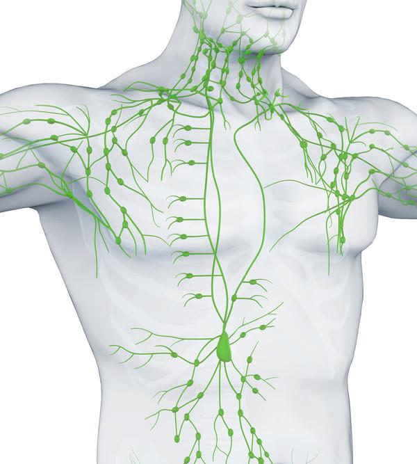
• 림프계. 암은 림프계로 전이되기 시작한 곳에서부터 퍼진다. 암은 림프관을 통해 몸의 다른 부분으로 이동한다.
• 혈액. 암은 혈액 속으로 들어가면서부터 퍼진다. 암은 혈관을 통해 신체의 다른 부분으로 이동한다.
암은 시작된 곳에서 신체의 다른 부분으로 전이될 수 있다. 암세포는 그들이 시작했던 곳에서 분리되어 림프계나 혈액을 통해 이동한다.
• 림프계. 암은 림프계에 들어가 림프관을 통과하며 몸의 다른 부분에 종양을 형성한다.
• 혈액. 암은 혈액 속으로 들어가 혈관을 통과하여 신체의 다른 부분에 종양(전이 암)을 형성한다.
전이성 종양은 1차 종양과 같은 종류의 종양이다. 예를 들어, 췌장 신경 내분비 종양이 간으로 퍼진다면, 간의 종양 세포는 실제로 신경 내분비 종양 세포다. 그 질병은 간암이 아닌 전이성 췌장 신경 내분비종양이다.
3. 재발 췌장 신경내분비종양
재발 췌장 신경 내분비 종양은 치료 후 재발한 종양이다. 종양은 췌장이나 신체의 다른 부분에 재발할 수 있다.
4. 치료 옵션의 개요
췌장 NET환자를 위한 치료에는 여러 종류가 있다. 췌장 신경 내분비종양(NET) 환자는 다양한 유형의 치료를 받을 수 있다. 일부 치료는 표준 치료(현재 사용되는 치료)이며, 일부는 임상 시험에서 테스트되고 있습니다. 치료 임상시험은 암환자에 대한 현재의 치료법을 개선하거나 새로운 치료법에 대한 정보를 얻는 것을 돕기 위한 연구이다. 임상 실험에서 새로운 치료법이 표준 치료법보다 나은 것으로 나타날 경우, 새로운 치료법이 표준 치료법이 될 수 있다. 환자들은 임상 실험에 참여하는 것에 대해 생각하고 싶어 할 수도 있다. 일부 임상 실험은 치료를 시작하지 않은 환자들에게만 개방된다. 6가지 유형의 표준 치료법이 사용된다.
수술
종양을 제거하기 위해 수술을 할 수도 있다. 다음 수술 유형 중 하나를 사용할 수 있습니다.
• 적출 : 종양만 제거하는 수술. 이것은 암이 췌장의 한 곳에서 발생할 때 수행될 수 있다.
• 췌십이지장 절제술 : 췌장의 머리, 쓸개, 근처의 림프절 및 위, 소장과 담관의 일부를 제거하는 수술. 췌장은 소화액과 인슐린을 만들기 위해 충분히 남겨둔다. 이 시술 중에 제거된 기관은 환자의 상태에 따라 달라집니다. 이를 Whipple 시술라고도 한다.
• 원위부 췌장절제술 : 췌장의 몸과 꼬리를 제거하는 수술. 암이 비장으로 전이된 경우에도 비장을 제거할 수 있다.
• 전체 위 절제술 : 위 전체를 제거하는 수술
• 위벽미주신경절단 : 위세포가 산을 만드는 신경을 자르는 수술
• 간 절제 : 간 일부 또는 전체를 제거하는 수술
• 고주파 절제 : 암세포를 죽이는 작은 전극을 부착한 특별한 탐침의 사용. 때로는 탐칩을 피부를 통해 직접 삽입하고 국소 마취만 하면 된다. 다른 경우에는 복부의 절개를 통해 탐침을 삽입한다. 이것은 병원에서 전신마취로 이루어진다.
• 냉동 외과적 절제술 : 비정상적인 세포를 파괴하기 위해 조직을 동결시키는 시술. 이것은 보통 액체 질소나 액체 이산화탄소를 포함하는 특별한 기구로 행해진다. 수술 또는 복강경 검사 중에 사용하거나 피부를 통해 삽입할 수 있다. 이 절차를 냉동 절제술이라고도 한다.
화학요법
화학요법은 암세포를 죽이거나 분열하는 것을 막음으로써 암세포의 성장을 막는데 약을 사용하는 암 치료법이다. 화학요법을 입으로 복용하거나 정맥이나 근육에 주입하면 혈류로 들어가 몸 전체에 걸쳐 암세포에 이를 수 있다. 화학요법이 뇌척수액, 장기 또는 복부와 같은 체강으로 직접 옮겨질 때, 약물은 주로 그러한 부위의 암세포에 영향을 미친다. 화학요법은 둘 이상의 항암제를 사용하는 것이다. 화학요법이 주어지는 방법은 치료되는 암의 종류에 따라 다르다.
호르몬 치료
호르몬 치료는 호르몬을 제거하거나 그들의 행동을 막고 암세포가 자라는 것을 막는 암 치료법이다. 호르몬은 몸 안의 분비선에 의해 만들어지고 혈류로 순환되는 물질이다. 일부 호르몬은 특정 암을 성장시킬 수 있다. 암세포에 호르몬이 부착할 수 있는 곳(수용체)이 있다는 검사 결과가 나오면 약물 수술 방사선 치료를 통해 호르몬의 생성을 줄이거나 활동을 차단한다.
간 동맥 폐색 또는 화학 색전
간 동맥 폐색은 간 동맥을 통해 간으로 가는 혈액의 흐름을 막거나 줄이기 위해 약물, 작은 입자 또는 다른 물질을 사용한다. 이것은 간에서 자라는 암세포를 죽이기 위해 행해진다. 종양이 자라기 위해 필요한 산소와 영양분을 얻는 것을 막는다. 간은 위와 장에서 혈액을 운반하는 간 문맥에서 혈액을 계속 받는다.
간 동맥 폐색 시 전달되는 화학요법은 화학색전술이라 불린다. 항암제는 카테터(얇은 튜브)를 통해 간 동맥에 주입된다. 그 약은 동맥을 막고 종양으로 가는 혈류를 차단하는 물질과 혼합되어 있다. 대부분의 항암제는 종양 근처에 갇혀 있고 소량의 항암제만이 신체의 다른 부분에 도달한다. 폐색은 동맥을 차단하는 데 사용되는 물질에 따라 일시적이거나 영구적일 수 있다.
표적치료
표적 치료는 약물이나 다른 물질을 사용하여 정상 세포를 해치지 않고 특정 암 세포를 식별하고 공격하는 치료의 한 종류이다. 특정 유형의 표적 치료법이 췌장 NET 치료에서 연구되고 있다.
지지요법
질병이나 치료로 인한 문제를 줄이기 위해 도움을 준다. 췌장 NET에 대한 지지요법에는 다음과 같은 치료법이 포함될 수 있습니다.
• 위궤양은 아래와 같은 약물로 치료할 수 있다
- 오메프라졸, 랜소프라졸 또는 판토프라졸과 같은 양자 펌프 억제제
- 시메티딘, 라니티딘, 파모티딘과 같은 약물을 차단하는 히스타민 억제제
- 옥트레오티드와 같은 소마토스타틴 형태의 약물
• 설사는 다음과 같이 치료할 수 있다
- 칼륨 또는 염화물과 같은 전해질을 함유한 정맥주입용 (IV) 수액
- 옥트레오티드와 같은 소마토스타틴 형태의 약물
• 저혈당은 소량의 식사를 자주 하거나 정상적인 혈당 수치를 유지하기 위한 약물 요법으로 치료할 수 있다.
• 고혈당은 구강 또는 인슐린 주사를 통해 복용하는 약물로 치료할 수 있다.
환자들은 임상 실험에 참여하는 것에 대해 생각할 수도 있다. 일부 환자의 경우 임상 실험에 참여하는 것이 최선의 치료 선택일 수 있다. 임상 실험은 암 연구 과정의 일부이다. 새로운 암 치료법이 안전하고 효과적이거나 표준 치료법보다 나은지 알아내기 위해 임상 실험이 행해진다.
오늘날 암에 대한 많은 표준 치료법은 이전의 임상 실험에 기초하고 있다. 임상 실험에 참여하는 환자는 표준 치료를 받거나 새로운 치료를 처음 받을 수 있다.
임상 실험에 참여하는 환자들은 또한 미래에 암이 치료될 방법을 개선하는데 도움을 준다. 임상 실험이 효과적인 새로운 치료로 이어지지 않을 때에도 그들은 종종 중요한 질문에 대답하고 연구를 진전시키는데 도움을 준다.
환자는 암 치료를 시작하기 전, 도중 또는 시작한 후에 임상 실험에 들어갈 수 있다. 일부 임상 실험에는 아직 치료를 받지 못한 환자만 포함된다. 암이 낫지 않은 환자에 대한 다른 시험 치료법. 또한 암이 재발하는 것을 막거나 암 치료의 부작용을 줄일 수 있는 새로운 방법을 시험하는 임상 실험도 있다.
임상 실험이 전국 많은 지역에서 일어나고 있다. NCI가 지원하는 임상시험에 대한 정보는 NCI의 임상시험 검색 웹페이지에서 확인할 수 있다. 다른 기관에서 지원하는 임상 시험은 ClinicalTrials.gov 웹사이트에서 볼 수 있다.
후속 추적검사가 필요할 수 있다
암을 진단하거나 암의 단계를 알아내기 위해 수행된 일부 테스트가 반복될 수 있습니다. 일부 테스트는 치료가 얼마나 잘 작동하는지 확인하기 위해 반복됩니다. 치료를 계속할지, 변경할지 또는 중지할지에 대한 결정은 이러한 테스트 결과에 기초할 수 있다.
일부 검사는 치료가 끝난 후에도 때때로 계속 수행될 것이다. 이 테스트의 결과는 상태가 바뀌었는지 또는 암이 재발했는지 알 수 있다. 이러한 테스트를 Follow up(팔로우업) 테스트라고도 한다.
4. 췌장 신경내분비종양의 치료방법
가스트린종
가스트린종의 치료에는 지지 및 다음 사항이 포함될 수 있다.
• 위산 과다로 인한 증상의 경우, 위산 함량을 감소시키는 약물이 될 수 있다.
• 췌두(췌장 머리)의 단일 종양
- 종양을 제거하는 수술
- 위세포가 산을 만드는 신경을 절단하는 수술, 위산을 감소시키는 약으로 치료
- 위 전체를 제거하는 수술(이런 경우는 희귀하다)
• 췌장의 체부(몸)나 미부(꼬리 부분)의 단일 종양의 경우, 치료는 일반적으로 췌장의 체부(몸)나 미부(꼬리 부분)를 제거하는 수술이다.
• 췌장에 있는 복수 종양의 경우, 치료는 일반적으로 췌장의 몸이나 꼬리를 제거하는 수술이다. 수술 후에도 종양이 남아 있는 경우 치료에는 다음 중 하나가 포함될 수 있다.
- 위세포가 산을 만드는 신경을 절단하는 수술 및 위산을 감소시키는 약물로 치료
- 위 전체를 제거하는 수술(희귀)
• 십이지장에서 하나 이상의 종양(위장에 연결된 소장의 일부)에 대한 치료는 대개 췌장두절제술(췌장두, 담낭, 근처 림프절 및 위, 소장, 담관 일부를 제거하는 수술)이다.
• 종양이 발견되지 않는 경우, 치료에는 다음이 포함될 수 있다.
- 위세포가 산을 만드는 신경을 절단하는 수술, 위산을 감소시키는 약으로 치료
- 위 전체를 제거하는 수술(희귀)
• 암이 간까지 전이된 경우 치료에는 다음이 포함될 수 있다.
- 간의 일부 또는 전체를 제거하는 수술
- 고주파 절제 또는 극저온 절제
- 화학색전술
• 암이 인체의 다른 부분으로 전이되었거나 위산을 감소시키기 위한 수술이나 약물 치료로 나아지지 않는 경우, 치료는 다음을 포함할 수 있다.
- 화학요법
- 호르몬요법
• 암이 대부분 간에 영향을 미치고 호르몬이나 종양 크기 때문에 환자가 심각한 증상을 보이는 경우 치료에는 다음이 포함될 수 있다.
- 전신 화학 요법의 유무에 관계없이 간 동맥 폐색
- 전신 화학 요법의 유무에 관계없이 화학색전
인슐린종
인슐린종 치료에는 다음이 포함될 수 있다.
• 췌장의 머리나 꼬리에 있는 하나의 작은 종양의 경우, 치료는 보통 종양을 제거하기 위한 수술이다.
• 수술로 제거할 수 없는 췌장 머리의 하나의 큰 종양의 경우, 치료는 대개 췌장 절제술(췌장 머리, 쓸개, 근처 림프절 및 위, 소장과 담관의 일부를 제거하는 수술)이다.
• 췌장의 몸이나 꼬리에 있는 하나의 큰 종양의 경우, 치료는 대개 원위부 췌장절제술(췌장의 몸과 꼬리를 제거하는 수술)이다.
• 췌장에 있는 하나 이상의 종양의 경우, 치료는 일반적으로 췌장의 머리와 몸통과 꼬리 부분의 종양을 제거하는 수술이다.
• 수술로 제거할 수 없는 종양의 경우, 치료에는 다음이 포함될 수 있다.
- 결합화학요법
- 췌장에 의한 인슐린의 양을 줄이기 위한 약물 치료
- 호르몬요법
- 고주파 절제 또는 극저온 절제.
• 림프절이나 신체의 다른 부분으로 전이된 암의 경우 치료에는 다음이 포함될 수 있다.
- 암 제거 수술
- 수술로 암을 제거할 수 없는 경우, 고주파 절제 또는 극저온 절제
• 암이 대부분 간에 영향을 미치고 호르몬이나 종양 크기 때문에 환자가 심각한 증상을 보이는 경우 치료에는 다음이 포함될 수 있습니다.
- 전신 화학 요법의 유무에 관계없이 간 동맥 폐색
- 전신 화학 요법의 유무에 관계없이 화학 색전
기타 췌장 신경 내분비 종양(섬 세포 종양)
VIPoma의 경우 시술에는 다음이 포함될 수 있다.
• 체내에서 손실된 체액과 전해질을 대체하기 위한 체액 및 호르몬 치료
• 종양 및 주변 림프절을 제거하는 수술
• 종양을 완전히 제거할 수 없거나 신체의 먼 곳으로 퍼진 경우 가능한 한 많은 종양을 제거하는 수술 이것은 증상을 완화시키고 삶의 질을 향상시키기 위한 완화요법이다.
• 림프절이나 신체의 다른 부분으로 전이된 종양의 경우, 치료에는 다음이 포함될 수 있다.
- 종양을 제거하는 수술
- 수술로 종양을 제거할 수 없는 경우, 고주파 절제 또는 극저온 절제
• 치료 중에 계속 성장하거나 신체의 다른 부분으로 전이된 종양의 경우, 치료에는 다음이 포함될 수 있다.
-화학요법
- 표적 치료
소마토스타틴종의 경우, 치료에는 다음이 포함될 수 있다
•종양을 제거하는 수술
• 신체의 먼 곳으로 전이된 암의 경우, 가능한 한 많은 암을 제거하여 증상을 완화하고 삶의 질을 향상시키는 수술
• 치료 중에 계속 성장하거나 신체의 다른 부분으로 전이된 종양의 경우, 치료에는 다음이 포함될 수 있다.
- 화학요법
- 표적요법
다른 유형의 췌장 신경 내분비 종양(NET) 치료에는 다음이 포함될 수 있다
• 종양을 제거하는 수술
• 몸 먼 곳으로 전이된 암의 경우 암을 최대한 제거하는 수술이나 증상을 완화하고 삶의 질을 높이기 위한 호르몬 요법
• 치료 중에 계속 성장하거나 신체의 다른 부분으로 전이된 종양의 경우, 치료에는 다음이 포함될 수 있다.
- 화학요법
- 표적요법
재발 또는 진행성 췌장 신경 내분비 종양(섬 세포 종양)
치료 중 또는 재발(돌아옴)하는 동안 계속 자라는 췌장 신경 내분비 종양(NET)의 치료에는 다음이 포함될 수 있다.
• 종양을 절제하는 수술
• 화학요법
• 호르몬 치료
• 표적 치료
• 간 전이의 경우
- 국소 화학 요법
- 전신 화학 요법의 유무에 관계없이 간 동맥 폐색 또는 화학색전
• 새로운 치료법의 임상 시험
당사의 임상 시험 검색을 사용하여 환자를 수용하는 NCI 지원 암 임상 시험을 찾다. 암의 유형, 환자의 나이 및 수행 위치를 기준으로 시행을 검색할 수 있다. 임상 시험에 대한 일반적인 정보도 이용할 수 있다.
Pancreatic Neuroendocrine Tumors (Islet Cell Tumors) Treatment (PDQ®)–Patient Version
Updated: February 17, 2020
On This Page
•General Information About Pancreatic Neuroendocrine Tumors (Islet Cell Tumors)
•Stages of Pancreatic Neuroendocrine Tumors
•Recurrent Pancreatic Neuroendocrine Tumors
•Treatment Option Overview
•Treatment Options for Pancreatic Neuroendocrine Tumors
1. General Information About Pancreatic Neuroendocrine Tumors (Islet Cell Tumors)
Key Points
•Pancreatic neuroendocrine tumors form in hormone-making cells (islet cells) of the pancreas.
•Pancreatic NETs may or may not cause signs or symptoms.
•There are different kinds of functional pancreatic NETs.
•Having certain syndromes can increase the risk of pancreatic NETs.
•Different types of pancreatic NETs have different signs and symptoms.
•Lab tests and imaging tests are used to detect (find) and diagnose pancreatic NETs.
•Other kinds of lab tests are used to check for the specific type of pancreatic NETs.
•Certain factors affect prognosis (chance of recovery) and treatment options.
Pancreatic neuroendocrine tumors form in hormone-making cells (islet cells) of the pancreas.
The pancreas is a gland about 6 inches long that is shaped like a thin pear lying on its side. The wider end of the pancreas is called the head, the middle section is called the body, and the narrow end is called the tail. The pancreas lies behind the stomach and in front of the spine.
There are two kinds of cells in the pancreas:
•Endocrine pancreas cells make several kinds of hormones (chemicals that control the actions of certain cells or organs in the body), such as insulin to control blood sugar. They cluster together in many small groups (islets) throughout the pancreas. Endocrine pancreas cells are also called islet cells or islets of Langerhans. Tumors that form in islet cells are called islet cell tumors, pancreatic endocrine tumors, or pancreatic neuroendocrine tumors (pancreatic NETs).
•Exocrine pancreas cells make enzymes that are released into the small intestine to help the body digest food. Most of the pancreas is made of ducts with small sacs at the end of the ducts, which are lined with exocrine cells.
This summary discusses islet cell tumors of the endocrine pancreas. See the PDQ summary on Pancreatic Cancer Treatment (Adult) for information on exocrine pancreatic cancer.
Pancreatic neuroendocrine tumors (NETs) may be benign (not cancer) or malignant (cancer). When pancreatic NETs are malignant, they are called pancreatic endocrine cancer or islet cell carcinoma.
Pancreatic NETs are much less common than pancreatic exocrine tumors and have a better prognosis.
Pancreatic NETs may or may not cause signs or symptoms.
Pancreatic NETs may be functional or nonfunctional:
•Functional tumors make extra amounts of hormones, such as gastrin, insulin, and glucagon, that cause signs and symptoms.
•Nonfunctional tumors do not make extra amounts of hormones. Signs and symptoms are caused by the tumor as it spreads and grows. Most nonfunctional tumors are malignant (cancer).
Most pancreatic NETs are functional tumors.
There are different kinds of functional pancreatic NETs.
Pancreatic NETs make different kinds of hormones such as gastrin, insulin, and glucagon. Functional pancreatic NETs include the following:
•Gastrinoma: A tumor that forms in cells that make gastrin. Gastrin is a hormone that causes the stomach to release an acid that helps digest food. Both gastrin and stomach acid are increased by gastrinomas. When increased stomach acid, stomach ulcers, and diarrhea are caused by a tumor that makes gastrin, it is called Zollinger-Ellison syndrome. A gastrinoma usually forms in the head of the pancreas and sometimes forms in the small intestine. Most gastrinomas are malignant (cancer).
•Insulinoma: A tumor that forms in cells that make insulin. Insulin is a hormone that controls the amount of glucose (sugar) in the blood. It moves glucose into the cells, where it can be used by the body for energy. Insulinomas are usually slow-growing tumors that rarely spread. An insulinoma forms in the head, body, or tail of the pancreas. Insulinomas are usually benign (not cancer).
•Glucagonoma: A tumor that forms in cells that make glucagon. Glucagon is a hormone that increases the amount of glucose in the blood. It causes the liver to break down glycogen. Too much glucagon causes hyperglycemia (high blood sugar). A glucagonoma usually forms in the tail of the pancreas. Most glucagonomas are malignant (cancer).
•Other types of tumors: There are other rare types of functional pancreatic NETs that make hormones, including hormones that control the balance of sugar, salt, and water in the body. These tumors include:
-VIPomas, which make vasoactive intestinal peptide. VIPoma may also be called Verner-Morrison syndrome.
-Somatostatinomas, which make somatostatin.
These other types of tumors are grouped together because they are treated in much the same way.
Having certain syndromes can increase the risk of pancreatic NETs.
Anything that increases your risk of getting a disease is called a risk factor. Having a risk factor does not mean that you will get cancer; not having risk factors doesn't mean that you will not get cancer. Talk with your doctor if you think you may be at risk.
Multiple endocrine neoplasia type 1 (MEN1) syndrome is a risk factor for pancreatic NETs.
Different types of pancreatic NETs have different signs and symptoms.
Signs or symptoms can be caused by the growth of the tumor and/or by hormones the tumor makes or by other conditions. Some tumors may not cause signs or symptoms. Check with your doctor if you have any of these problems.
Signs and symptoms of a non-functional pancreatic NET
A non-functional pancreatic NET may grow for a long time without causing signs or symptoms. It may grow large or spread to other parts of the body before it causes signs or symptoms, such as:
•Diarrhea.
•Indigestion.
•A lump in the abdomen.
•Pain in the abdomen or back.
•Yellowing of the skin and whites of the eyes.
Signs and symptoms of a functional pancreatic NET
The signs and symptoms of a functional pancreatic NET depend on the type of hormone being made.
Too much gastrin may cause:
•Stomach ulcers that keep coming back.
•Pain in the abdomen, which may spread to the back. The pain may come and go and it may go away after taking an antacid.
•The flow of stomach contents back into the esophagus (gastroesophageal reflux).
•Diarrhea.
Too much insulin may cause:
•Low blood sugar. This can cause blurred vision, headache, and feeling lightheaded, tired, weak, shaky, nervous, irritable, sweaty, confused, or hungry.
•Fast heartbeat.
Too much glucagon may cause:
•Skin rash on the face, stomach, or legs.
•High blood sugar. This can cause headaches, frequent urination, dry skin and mouth, or feeling hungry, thirsty, tired, or weak.
•Blood clots. Blood clots in the lung can cause shortness of breath, cough, or pain in the chest. Blood clots in the arm or leg can cause pain, swelling, warmth, or redness of the arm or leg.
•Diarrhea.
•Weight loss for no known reason.
•Sore tongue or sores at the corners of the mouth.
Too much vasoactive intestinal peptide (VIP) may cause:
•Very large amounts of watery diarrhea.
•Dehydration. This can cause feeling thirsty, making less urine, dry skin and mouth, headaches, dizziness, or feeling tired.
•Low potassium level in the blood. This can cause muscle weakness, aching, or cramps, numbness and tingling, frequent urination, fast heartbeat, and feeling confused or thirsty.
•Cramps or pain in the abdomen.
•Weight loss for no known reason.
Too much somatostatin may cause:
•High blood sugar. This can cause headaches, frequent urination, dry skin and mouth, or feeling hungry, thirsty, tired, or weak.
•Diarrhea.
•Steatorrhea (very foul-smelling stool that floats).
•Gallstones.
•Yellowing of the skin and whites of the eyes.
•Weight loss for no known reason.
A pancreatic NET may also make too much adrenocorticotropic hormone (ACTH) and cause Cushing syndrome. Signs and symptoms of Cushing syndrome include the following:
•Headache.
•Some loss of vision.
•Weight gain in the face, neck, and trunk of the body, and thin arms and legs.
•A lump of fat on the back of the neck.
•Thin skin that may have purple or pink stretch marks on the chest or abdomen.
•Easy bruising.
•Growth of fine hair on the face, upper back, or arms.
•Bones that break easily.
•Sores or cuts that heal slowly.
•Anxiety, irritability, and depression.
The treatment of pancreatic NETs that make too much ACTH and Cushing syndrome are not discussed in this summary.
Lab tests and imaging tests are used to detect (find) and diagnose pancreatic NETs.
The following tests and procedures may be used:
•Physical exam and history: An exam of the body to check general signs of health, including checking for signs of disease, such as lumps or anything else that seems unusual. A history of the patient’s health habits and past illnesses and treatments will also be taken.
•Blood chemistry studies: A procedure in which a blood sample is checked to measure the amounts of certain substances, such as glucose (sugar), released into the blood by organs and tissues in the body. An unusual (higher or lower than normal) amount of a substance can be a sign of disease.
•Chromogranin A test: A test in which a blood sample is checked to measure the amount of chromogranin A in the blood. A higher than normal amount of chromogranin A and normal amounts of hormones such as gastrin, insulin, and glucagon can be a sign of a non-functional pancreatic NET.
•Abdominal CT scan (CAT scan): A procedure that makes a series of detailed pictures of the abdomen, taken from different angles. The pictures are made by a computer linked to an x-ray machine. A dye may be injected into a vein or swallowed to help the organs or tissues show up more clearly. This procedure is also called computed tomography, computerized tomography, or computerized axial tomography.
•MRI (magnetic resonance imaging): A procedure that uses a magnet, radio waves, and a computer to make a series of detailed pictures of areas inside the body. This procedure is also called nuclear magnetic resonance imaging (NMRI).
•Somatostatin receptor scintigraphy: A type of radionuclide scan that may be used to find small pancreatic NETs. A small amount of radioactive octreotide (a hormone that attaches to tumors) is injected into a vein and travels through the blood. The radioactive octreotide attaches to the tumor and a special camera that detects radioactivity is used to show where the tumors are in the body. This procedure is also called octreotide scan and SRS.
•Endoscopic ultrasound (EUS): A procedure in which an endoscope is inserted into the body, usually through the mouth or rectum. An endoscope is a thin, tube-like instrument with a light and a lens for viewing. A probe at the end of the endoscope is used to bounce high-energy sound waves (ultrasound) off internal tissues or organs and make echoes. The echoes form a picture of body tissues called a sonogram. This procedure is also called endosonography.
•Endoscopic retrograde cholangiopancreatography (ERCP): A procedure used to x-ray the ducts (tubes) that carry bile from the liver to the gallbladder and from the gallbladder to the small intestine. Sometimes pancreatic cancer causes these ducts to narrow and block or slow the flow of bile, causing jaundice. An endoscope is passed through the mouth, esophagus, and stomach into the first part of the small intestine. An endoscope is a thin, tube-like instrument with a light and a lens for viewing. A catheter (a smaller tube) is then inserted through the endoscope into the pancreatic ducts. A dye is injected through the catheter into the ducts and an x-ray is taken. If the ducts are blocked by a tumor, a fine tube may be inserted into the duct to unblock it. This tube (or stent) may be left in place to keep the duct open. Tissue samples may also be taken and checked under a microscope for signs of cancer.
•Angiogram: A procedure to look at blood vessels and the flow of blood. A contrast dye is injected into the blood vessel. As the contrast dye moves through the blood vessel, x-rays are taken to see if there are any blockages.
•Laparotomy: A surgical procedure in which an incision (cut) is made in the wall of the abdomen to check the inside of the abdomen for signs of disease. The size of the incision depends on the reason the laparotomy is being done. Sometimes organs are removed or tissue samples are taken and checked under a microscope for signs of disease.
•Intraoperative ultrasound: A procedure that uses high-energy sound waves (ultrasound) to create images of internal organs or tissues during surgery. A transducer placed directly on the organ or tissue is used to make the sound waves, which create echoes. The transducer receives the echoes and sends them to a computer, which uses the echoes to make pictures called sonograms.
•Biopsy: The removal of cells or tissues so they can be viewed under a microscope by a pathologist to check for signs of cancer. There are several ways to do a biopsy for pancreatic NETs. Cells may be removed using a fine or wide needle inserted into the pancreas during an x-ray or ultrasound. Tissue may also be removed during a laparoscopy (a surgical incision made in the wall of the abdomen).
•Bone scan: A procedure to check if there are rapidly dividing cells, such as cancer cells, in the bone. A very small amount of radioactive material is injected into a vein and travels through the bloodstream. The radioactive material collects in bones with cancer and is detected by a scanner.
Other kinds of lab tests are used to check for the specific type of pancreatic NETs.
The following tests and procedures may be used:
Gastrinoma
•Fasting serum gastrin test: A test in which a blood sample is checked to measure the amount of gastrin in the blood. This test is done after the patient has had nothing to eat or drink for at least 8 hours. Conditions other than gastrinoma can cause an increase in the amount of gastrin in the blood.
•Basal acid output test: A test to measure the amount of acid made by the stomach. The test is done after the patient has had nothing to eat or drink for at least 8 hours. A tube is inserted through the nose or throat, into the stomach. The stomach contents are removed and four samples of gastric acid are removed through the tube. These samples are used to find out the amount of gastric acid made during the test and the pH level of the gastric secretions.
•Secretin stimulation test: If the basal acid output test result is not normal, a secretin stimulation test may be done. The tube is moved into the small intestine and samples are taken from the small intestine after a drug called secretin is injected. Secretin causes the small intestine to make acid. When there is a gastrinoma, the secretin causes an increase in how much gastric acid is made and the level of gastrin in the blood.
•Somatostatin receptor scintigraphy: A type of radionuclide scan that may be used to find small pancreatic NETs. A small amount of radioactive octreotide (a hormone that attaches to tumors) is injected into a vein and travels through the blood. The radioactive octreotide attaches to the tumor and a special camera that detects radioactivity is used to show where the tumors are in the body. This procedure is also called octreotide scan and SRS.
Insulinoma
•Fasting serum glucose and insulin test: A test in which a blood sample is checked to measure the amounts of glucose (sugar) and insulin in the blood. The test is done after the patient has had nothing to eat or drink for at least 24 hours.
Glucagonoma
•Fasting serum glucagon test: A test in which a blood sample is checked to measure the amount of glucagon in the blood. The test is done after the patient has had nothing to eat or drink for at least 8 hours.
Other tumor types
•VIPoma
•Serum VIP (vasoactive intestinal peptide) test: A test in which a blood sample is checked to measure the amount of VIP.
•Blood chemistry studies: A procedure in which a blood sample is checked to measure the amounts of certain substances released into the blood by organs and tissues in the body. An unusual (higher or lower than normal) amount of a substance can be a sign of disease. In VIPoma, there is a lower than normal amount of potassium.
•Stool analysis: A stool sample is checked for a higher than normal sodium (salt) and potassium levels.
•Somatostatinoma
•Fasting serum somatostatin test: A test in which a blood sample is checked to measure the amount of somatostatin in the blood. The test is done after the patient has had nothing to eat or drink for at least 8 hours.
•Somatostatin receptor scintigraphy: A type of radionuclide scan that may be used to find small pancreatic NETs. A small amount of radioactive octreotide (a hormone that attaches to tumors) is injected into a vein and travels through the blood. The radioactive octreotide attaches to the tumor and a special camera that detects radioactivity is used to show where the tumors are in the body. This procedure is also called octreotide scan and SRS.
Certain factors affect prognosis (chance of recovery) and treatment options.
Pancreatic NETs can often be cured. The prognosis (chance of recovery) and treatment options depend on the following:
•The type of cancer cell.
•Where the tumor is found in the pancreas.
•Whether the tumor has spread to more than one place in the pancreas or to other parts of the body.
•Whether the patient has MEN1 syndrome.
•The patient's age and general health.
•Whether the cancer has just been diagnosed or has recurred (come back).
2. Stages of Pancreatic Neuroendocrine Tumors
Key Points
•The plan for cancer treatment depends on where the NET is found in the pancreas and whether it has spread.
•There are three ways that cancer spreads in the body.
•Cancer may spread from where it began to other parts of the body.
The plan for cancer treatment depends on where the NET is found in the pancreas and whether it has spread.
The process used to find out if cancer has spread within the pancreas or to other parts of the body is called staging. The results of the tests and procedures used to diagnose pancreatic neuroendocrine tumors (NETs) are also used to find out whether the cancer has spread. See the General Information section for a description of these tests and procedures.
Although there is a standard staging system for pancreatic NETs, it is not used to plan treatment. Treatment of pancreatic NETs is based on the following:
•Whether the cancer is found in one place in the pancreas.
•Whether the cancer is found in several places in the pancreas.
•Whether the cancer has spread to lymph nodes near the pancreas or to other parts of the body such as the liver, lung, peritoneum, or bone.
There are three ways that cancer spreads in the body.
Cancer can spread through tissue, the lymph system, and the blood:
•Tissue. The cancer spreads from where it began by growing into nearby areas.
•Lymph system. The cancer spreads from where it began by getting into the lymph system. The cancer travels through the lymph vessels to other parts of the body.
•Blood. The cancer spreads from where it began by getting into the blood. The cancer travels through the blood vessels to other parts of the body.
Cancer may spread from where it began to other parts of the body.
When cancer spreads to another part of the body, it is called metastasis. Cancer cells break away from where they began (the primary tumor) and travel through the lymph system or blood.
•Lymph system. The cancer gets into the lymph system, travels through the lymph vessels, and forms a tumor (metastatic tumor) in another part of the body.
•Blood. The cancer gets into the blood, travels through the blood vessels, and forms a tumor (metastatic tumor) in another part of the body.
The metastatic tumor is the same type of tumor as the primary tumor. For example, if a pancreatic neuroendocrine tumor spreads to the liver, the tumor cells in the liver are actually neuroendocrine tumor cells. The disease is metastatic pancreatic neuroendocrine tumor, not liver cancer.
3. Recurrent Pancreatic Neuroendocrine Tumors
Recurrent pancreatic neuroendocrine tumors (NETs) are tumors that have recurred (come back) after being treated. The tumors may come back in the pancreas or in other parts of the body.
4. Treatment Option Overview
Key Points
•There are different types of treatment for patients with pancreatic NETs.
•Six types of standard treatment are used:
-Surgery
-Chemotherapy
-Hormone therapy
-Hepatic arterial occlusion or chemoembolization
-Targeted therapy
-Supportive care
•New types of treatment are being tested in clinical trials.
•Treatment for pancreatic neuroendocrine tumors may cause side effects.
•Patients may want to think about taking part in a clinical trial.
•Patients can enter clinical trials before, during, or after starting their cancer treatment.
•Follow-up tests may be needed.
There are different types of treatment for patients with pancreatic NETs.
Different types of treatments are available for patients with pancreatic neuroendocrine tumors (NETs). Some treatments are standard (the currently used treatment), and some are being tested in clinical trials. A treatment clinical trial is a research study meant to help improve current treatments or obtain information on new treatments for patients with cancer. When clinical trials show that a new treatment is better than the standard treatment, the new treatment may become the standard treatment. Patients may want to think about taking part in a clinical trial. Some clinical trials are open only to patients who have not started treatment.
Six types of standard treatment are used:
Surgery
An operation may be done to remove the tumor. One of the following types of surgery may be used:
•Enucleation: Surgery to remove the tumor only. This may be done when cancer occurs in one place in the pancreas.
•Pancreatoduodenectomy: A surgical procedure in which the head of the pancreas, the gallbladder, nearby lymph nodes and part of the stomach, small intestine, and bile duct are removed. Enough of the pancreas is left to make digestive juices and insulin. The organs removed during this procedure depend on the patient's condition. This is also called the Whipple procedure.
•Distal pancreatectomy: Surgery to remove the body and tail of the pancreas. The spleen may also be removed if cancer has spread to the spleen.
•Total gastrectomy: Surgery to remove the whole stomach.
•Parietal cell vagotomy: Surgery to cut the nerve that causes stomach cells to make acid.
•Liver resection: Surgery to remove part or all of the liver.
•Radiofrequency ablation: The use of a special probe with tiny electrodes that kill cancer cells. Sometimes the probe is inserted directly through the skin and only local anesthesia is needed. In other cases, the probe is inserted through an incision in the abdomen. This is done in the hospital with general anesthesia.
•Cryosurgical ablation: A procedure in which tissue is frozen to destroy abnormal cells. This is usually done with a special instrument that contains liquid nitrogen or liquid carbon dioxide. The instrument may be used during surgery or laparoscopy or inserted through the skin. This procedure is also called cryoablation.
Chemotherapy
Chemotherapy is a cancer treatment that uses drugs to stop the growth of cancer cells, either by killing the cells or by stopping them from dividing. When chemotherapy is taken by mouth or injected into a vein or muscle, the drugs enter the bloodstream and can reach cancer cells throughout the body (systemic chemotherapy). When chemotherapy is placed directly into the cerebrospinal fluid, an organ, or a body cavity such as the abdomen, the drugs mainly affect cancer cells in those areas (regional chemotherapy). Combination chemotherapy is the use of more than one anticancer drug. The way the chemotherapy is given depends on the type of the cancer being treated.
Hormone therapy
Hormone therapy is a cancer treatment that removes hormones or blocks their action and stops cancer cells from growing. Hormones are substances made by glands in the body and circulated in the bloodstream. Some hormones can cause certain cancers to grow. If tests show that the cancer cells have places where hormones can attach (receptors), drugs, surgery, or radiation therapy is used to reduce the production of hormones or block them from working.
Hepatic arterial occlusion or chemoembolization
Hepatic arterial occlusion uses drugs, small particles, or other agents to block or reduce the flow of blood to the liver through the hepatic artery (the major blood vessel that carries blood to the liver). This is done to kill cancer cells growing in the liver. The tumor is prevented from getting the oxygen and nutrients it needs to grow. The liver continues to receive blood from the hepatic portal vein, which carries blood from the stomach and intestine.
Chemotherapy delivered during hepatic arterial occlusion is called chemoembolization. The anticancer drug is injected into the hepatic artery through a catheter (thin tube). The drug is mixed with the substance that blocks the artery and cuts off blood flow to the tumor. Most of the anticancer drug is trapped near the tumor and only a small amount of the drug reaches other parts of the body.
The blockage may be temporary or permanent, depending on the substance used to block the artery.
Targeted therapy
Targeted therapy is a type of treatment that uses drugs or other substances to identify and attack specific cancer cells without harming normal cells. Certain types of targeted therapies are being studied in the treatment of pancreatic NETs.
Supportive care
Supportive care is given to lessen the problems caused by the disease or its treatment. Supportive care for pancreatic NETs may include treatment for the following:
•Stomach ulcers may be treated with drug therapy such as:
•Proton pump inhibitor drugs such as omeprazole, lansoprazole, or pantoprazole.
•Histamine blocking drugs such as cimetidine, ranitidine, or famotidine.
•Somatostatin-type drugs such as octreotide.
•Diarrhea may be treated with:
•Intravenous (IV) fluids with electrolytes such as potassium or chloride.
•Somatostatin-type drugs such as octreotide.
•Low blood sugar may be treated by having small, frequent meals or with drug therapy to maintain a normal blood sugar level.
•High blood sugar may be treated with drugs taken by mouth or insulin by injection.
New types of treatment are being tested in clinical trials.
Information about clinical trials is available from the NCI website.
Treatment for pancreatic neuroendocrine tumors may cause side effects.
For information about side effects caused by treatment for cancer, see our Side Effects page.
Patients may want to think about taking part in a clinical trial.
For some patients, taking part in a clinical trial may be the best treatment choice. Clinical trials are part of the cancer research process. Clinical trials are done to find out if new cancer treatments are safe and effective or better than the standard treatment.
Many of today's standard treatments for cancer are based on earlier clinical trials. Patients who take part in a clinical trial may receive the standard treatment or be among the first to receive a new treatment.
Patients who take part in clinical trials also help improve the way cancer will be treated in the future. Even when clinical trials do not lead to effective new treatments, they often answer important questions and help move research forward.
Patients can enter clinical trials before, during, or after starting their cancer treatment.
Some clinical trials only include patients who have not yet received treatment. Other trials test treatments for patients whose cancer has not gotten better. There are also clinical trials that test new ways to stop cancer from recurring (coming back) or reduce the side effects of cancer treatment.
Clinical trials are taking place in many parts of the country. Information about clinical trials supported by NCI can be found on NCI’s clinical trials search webpage. Clinical trials supported by other organizations can be found on the ClinicalTrials.gov website.
Follow-up tests may be needed.
Some of the tests that were done to diagnose the cancer or to find out the stage of the cancer may be repeated. Some tests will be repeated in order to see how well the treatment is working. Decisions about whether to continue, change, or stop treatment may be based on the results of these tests.
Some of the tests will continue to be done from time to time after treatment has ended. The results of these tests can show if your condition has changed or if the cancer has recurred (come back). These tests are sometimes called follow-up tests or check-ups.
4. Treatment Options for Pancreatic Neuroendocrine Tumors
In This Section
•Gastrinoma
•Insulinoma
•Glucagonoma
•Other Pancreatic Neuroendocrine Tumors (Islet Cell Tumors)
•Recurrent or Progressive Pancreatic Neuroendocrine Tumors (Islet Cell Tumors)
For information about the treatments listed below, see the Treatment Option Overview section.
Gastrinoma
Treatment of gastrinoma may include supportive care and the following:
•For symptoms caused by too much stomach acid, treatment may be a drug that decreases the amount of acid made by the stomach.
•For a single tumor in the head of the pancreas:
-Surgery to remove the tumor.
-Surgery to cut the nerve that causes stomach cells to make acid and treatment with a drug that decreases stomach acid.
-Surgery to remove the whole stomach (rare).
•For a single tumor in the body or tail of the pancreas, treatment is usually surgery to remove the body or tail of the pancreas.
•For several tumors in the pancreas, treatment is usually surgery to remove the body or tail of the pancreas. If tumor remains after surgery, treatment may include either:
-Surgery to cut the nerve that causes stomach cells to make acid and treatment with a drug that decreases stomach acid; or
-Surgery to remove the whole stomach (rare).
•For one or more tumors in the duodenum (the part of the small intestine that connects to the stomach), treatment is usually pancreatoduodenectomy (surgery to remove the head of the pancreas, the gallbladder, nearby lymph nodes and part of the stomach, small intestine, and bile duct).
•If no tumor is found, treatment may include the following:
-Surgery to cut the nerve that causes stomach cells to make acid and treatment with a drug that decreases stomach acid.
-Surgery to remove the whole stomach (rare).
•If the cancer has spread to the liver, treatment may include:
-Surgery to remove part or all of the liver.
-Radiofrequency ablation or cryosurgical ablation.
-Chemoembolization.
•If cancer has spread to other parts of the body or does not get better with surgery or drugs to decrease stomach acid, treatment may include:
-Chemotherapy.
-Hormone therapy.
•If the cancer mostly affects the liver and the patient has severe symptoms from hormones or from the size of tumor, treatment may include:
-Hepatic arterial occlusion, with or without systemic chemotherapy.
-Chemoembolization, with or without systemic chemotherapy.
Use our clinical trial search to find NCI-supported cancer clinical trials that are accepting patients. You can search for trials based on the type of cancer, the age of the patient, and where the trials are being done. General information about clinical trials is also available.
Insulinoma
Treatment of insulinoma may include the following:
•For one small tumor in the head or tail of the pancreas, treatment is usually surgery to remove the tumor.
•For one large tumor in the head of the pancreas that cannot be removed by surgery, treatment is usually pancreatoduodenectomy (surgery to remove the head of the pancreas, the gallbladder, nearby lymph nodes and part of the stomach, small intestine, and bile duct).
•For one large tumor in the body or tail of the pancreas, treatment is usually a distal pancreatectomy (surgery to remove the body and tail of the pancreas).
•For more than one tumor in the pancreas, treatment is usually surgery to remove any tumors in the head of the pancreas and the body and tail of the pancreas.
•For tumors that cannot be removed by surgery, treatment may include the following:
-Combination chemotherapy.
-Palliative drug therapy to decrease the amount of insulin made by the pancreas.
-Hormone therapy.
-Radiofrequency ablation or cryosurgical ablation.
•For cancer that has spread to lymph nodes or other parts of the body, treatment may include the following:
-Surgery to remove the cancer.
-Radiofrequency ablation or cryosurgical ablation, if the cancer cannot be removed by surgery.
•If the cancer mostly affects the liver and the patient has severe symptoms from hormones or from the size of tumor, treatment may include:
-Hepatic arterial occlusion, with or without systemic chemotherapy.
-Chemoembolization, with or without systemic chemotherapy.
Use our clinical trial search to find NCI-supported cancer clinical trials that are accepting patients. You can search for trials based on the type of cancer, the age of the patient, and where the trials are being done. General information about clinical trials is also available.
Glucagonoma
Treatment may include the following:
•For one small tumor in the head or tail of the pancreas, treatment is usually surgery to remove the tumor.
•For one large tumor in the head of the pancreas that cannot be removed by surgery, treatment is usually pancreatoduodenectomy (surgery to remove the head of the pancreas, the gallbladder, nearby lymph nodes and part of the stomach, small intestine, and bile duct).
•For more than one tumor in the pancreas, treatment is usually surgery to remove the tumor or surgery to remove the body and tail of the pancreas.
•For tumors that cannot be removed by surgery, treatment may include the following:
-Combination chemotherapy.
-Hormone therapy.
-Radiofrequency ablation or cryosurgical ablation.
•For cancer that has spread to lymph nodes or other parts of the body, treatment may include the following:
-Surgery to remove the cancer.
-Radiofrequency ablation or cryosurgical ablation, if the cancer cannot be removed by surgery.
•If the cancer mostly affects the liver and the patient has severe symptoms from hormones or from the size of tumor, treatment may include:
-Hepatic arterial occlusion, with or without systemic chemotherapy.
-Chemoembolization, with or without systemic chemotherapy.
Use our clinical trial search to find NCI-supported cancer clinical trials that are accepting patients. You can search for trials based on the type of cancer, the age of the patient, and where the trials are being done. General information about clinical trials is also available.
Other Pancreatic Neuroendocrine Tumors (Islet Cell Tumors)
For VIPoma, treatment may include the following:
•Fluids and hormone therapy to replace fluids and electrolytes that have been lost from the body.
•Surgery to remove the tumor and nearby lymph nodes.
•Surgery to remove as much of the tumor as possible when the tumor cannot be completely removed or has spread to distant parts of the body. This is palliative therapy to relieve symptoms and improve the quality of life.
•For tumors that have spread to lymph nodes or other parts of the body, treatment may include the following:
-Surgery to remove the tumor.
-Radiofrequency ablation or cryosurgical ablation, if the tumor cannot be removed by surgery.
•For tumors that continue to grow during treatment or have spread to other parts of the body, treatment may include the following:
-Chemotherapy.
-Targeted therapy.
For somatostatinoma, treatment may include the following:
•Surgery to remove the tumor.
•For cancer that has spread to distant parts of the body, surgery to remove as much of the cancer as possible to relieve symptoms and improve quality of life.
•For tumors that continue to grow during treatment or have spread to other parts of the body, treatment may include the following:
-Chemotherapy.
-Targeted therapy.
Treatment of other types of pancreatic neuroendocrine tumors (NETs) may include the following:
•Surgery to remove the tumor.
•For cancer that has spread to distant parts of the body, surgery to remove as much of the cancer as possible or hormone therapy to relieve symptoms and improve quality of life.
•For tumors that continue to grow during treatment or have spread to other parts of the body, treatment may include the following:
-Chemotherapy.
-Targeted therapy.
Use our clinical trial search to find NCI-supported cancer clinical trials that are accepting patients. You can search for trials based on the type of cancer, the age of the patient, and where the trials are being done. General information about clinical trials is also available.
Recurrent or Progressive Pancreatic Neuroendocrine Tumors (Islet Cell Tumors)
Treatment of pancreatic neuroendocrine tumors (NETs) that continue to grow during treatment or recur (come back) may include the following:
• Surgery to remove the tumor.
•Chemotherapy.
•Hormone therapy.
•Targeted therapy.
•For liver metastases:
-Regional chemotherapy.
-Hepatic arterial occlusion or chemoembolization, with or without systemic chemotherapy.
•A clinical trial of a new therapy.
Use our clinical trial search to find NCI-supported cancer clinical trials that are accepting patients. You can search for trials based on the type of cancer, the age of the patient, and where the trials are being done. General information about clinical trials is also available.
암스쿨에 게재된 기사는 미국국립암연구소(NCI), 미국암협회(ACS), 국립암센터(NCC), 일본국립암연구소(NCCJ), 엠디앤더슨암센터(MD Anderson Cancer Center) 등 검증된 기관의 검증된 자료를 토대로 작성되었습니다.
이 글은 2021년 1월 작성 시점의 정보입니다. 치료 기준·급여 정보는 바뀌었을 수 있으니, 진료와 치료 결정은 반드시 담당 의료진과 상의하세요.