중추신경계 종양 치료 종합정보
2021.01.08 · 전영조 전문위원

성인 중추신경계 종양에 대한 일반 정보
성인 중추신경계 종양은 뇌 또는 척수의 조직에서 비정상적인 세포가 형성되는 질병이다. 뇌종양과 척수종양에는 많은 종류가 있다. 종양은 세포의 비정상적인 성장에 의해 형성되며 뇌나 척수의 다른 부분에서 시작될 수 있다. 뇌와 척수는 함께 중추신경계를 구성한다.
종양은 양성 또는 악성일 수 있다.
• 양성 뇌와 척수 종양이 성장하여 뇌 근처를 압박한다. 그것들은 다른 조직으로 거의 퍼지지 않고 재발할 수 있다.
• 악성 뇌와 척수 종양은 빠르게 성장하여 다른 뇌 조직으로 퍼질 수 있다.
종양이 뇌의 한 부분으로 자라거나 압박할 때, 그것은 뇌의 그 부분이 제대로 작용하는 것을 멈출 수 있다. 양성 뇌종양과 악성 뇌종양은 모두 증상과 징후를 유발하며 치료가 필요하다. 뇌와 척수 종양은 성인과 어린이 모두에게 발생할 수 있다. 그러나 어린이를 위한 치료는 어른을 위한 치료와 다를 수 있다.
신체의 다른 부분에서 시작해서 뇌로 퍼지는 종양을 전이성 뇌종양이라고 부른다. 뇌에서 시작되는 종양은 1차 뇌종양이라고 불린다. 1차 뇌종양은 뇌의 다른 부분이나 척추로 퍼질 수 있다. 그것들은 몸의 다른 부분으로 거의 퍼지지 않는다.
종종, 뇌에서 발견되는 종양은 신체의 다른 곳에서 시작되어 뇌의 한 곳 이상의 부분으로 퍼진다. 이것들은 전이성 뇌종양이라고 불린다. 전이성 뇌종양은 1차 뇌종양보다 더 흔하다. 전이성 뇌종양의 절반 이상은 폐암에 기인한 것이다. 일반적으로 뇌로 전이되는 다른 유형의 암은 다음과 같다.
• 흑색종
• 유방암
• 대장암
• 신장암
• 비인두암
• 일차 부위(원발소) 불명의 암
암은 뇌와 척수를 덮고 있는 두 개의 가장 안쪽 막으로 전이될 수 있다. 이것은 연수막 암종증이라고 불린다. 연수막으로 전이되는 가장 일반적인 암은 다음과 같다.
• 유방암
• 폐암
• 백혈병
• 림프종
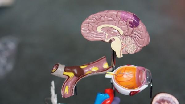
뇌는 많은 중요한 신체 기능을 통제한다. 뇌는 세 가지 주요 부분으로 이루어져 있다.
• 대뇌는 뇌의 가장 큰 부분이다. 그것은 머리 꼭대기에 있다. 대뇌는 사고와 학습, 문제 해결, 감정, 언어, 읽기, 쓰기, 그리고 자발적인 움직임을 통제한다.
• 소뇌는 뇌의 뒤 아래쪽에 있고(두부의 후면 가운데 가까운 쪽), 동작과 균형 및 자세를 관장한다.
• 뇌간은 뇌를 척수와 연결한다. 그것은 뇌의 가장 낮은 부분(목 바로 뒷부분 위)에 있다. 뇌간은 호흡과 심박수, 보고, 듣고, 걷고, 말하고, 먹는데 사용되는 신경과 근육을 조절한다.
척수는 뇌와 신체의 대부분의 신경들을 연결시킨다
척수는 뇌간에서 등 중앙을 따라 이어지는 신경 조직의 기둥이다. 그것은 막이라고 불리는 세 개의 얇은 조직의 층으로 덮여 있다. 이 막들은 척추 뼈로 둘러싸여 있다. 척수 신경은 근육을 움직이게 하는 뇌로부터의 메시지나 피부에서 뇌로 촉각을 느끼도록 하는 메시지와 같은 뇌와 신체의 나머지 부분 사이에 메시지를 전달한다.
뇌종양과 척수종양은 여러 종류가 있다
뇌와 척수 종양은 그들이 형성한 세포의 종류와 중추신경계(CNS)에서 종양이 처음 형성된 곳을 기준으로 이름 지어진다. 종양의 등급은 천천히 자라는 유형과 빠르게 성장하는 유형의 종양을 구별하는 데 사용될 수 있다. 세계보건기구(WHO)의 종양 등급은 암세포가 현미경 아래 얼마나 비정상적으로 보이는지, 그리고 얼마나 빨리 종양이 자라고 퍼질 수 있는지를 기준으로 한다.
세계보건기구 종양등급제
• 1등급 (저 등급) - 종양 세포는 현미경으로 보면 정상 세포와 더 비슷하며 II, III, IV급 종양 세포보다 더 느리게 성장하고 퍼져나간다. 그것들은 인근 조직으로 퍼지는 경우는 거의 없다. 만약 수술로 완전히 제거된다면 1급 뇌종양은 치료될 수 있다.
• II등급 - 종양 세포는 III등급 및 IV등급 종양 세포보다 더 느리게 성장하고 퍼진다. 그것들은 인근 조직으로 퍼져서 재발할 수 있다. 어떤 종양은 고 등급의 종양이 될 수도 있다.
• III등급 - 종양 세포는 현미경으로 보면 정상 세포와 매우 다르게 보이고 I등급 및 II등급 종양 세포보다 더 빨리 성장한다. 그것들은 근처 조직으로 퍼질 가능성이 있다.
• IV등급(고등급 ) - 종양 세포는 현미경으로 보면 정상 세포처럼 보이지 않으며 매우 빠르게 성장하고 퍼져나간다. 종양에는 죽은 세포가 있을 수 있다. 4급 종양은 보통 완치(치유)할 수 없다.
뇌 또는 척수에 다음과 같은 유형의 1차 종양이 형성될 수 있다
성상 교종
성상 세포종은 성상세포라는 별 모양의 뇌세포에서 시작되는데, 이것은 신경세포를 건강하게 유지하는데 도움을 준다. 성상세포는 교(질)세포의 한 종류이다. 교질세포는 때때로 신경교종이라는 종양을 형성한다. 성상 교종에는 다음과 같은 종양이 포함된다.
• 뇌간 교증(일반적으로 높은 등급) : 뇌간 교증은 척수와 연결된 뇌의 한 부분인 뇌간에서 형성된다. 그것은 종종 뇌간을 통해 널리 퍼지고 치료하기 어려운 높은 등급의 종양이다. 뇌간 교종은 성인에게서 드물다.
• 송과체 성상교종(모든 등급) : 송과체 성상세포종은 송과선 주변의 조직에서 형성되며 어떠한 등급도 될 수 있다. 송과선은 수면과 깨어있는(각성) 주기를 조절하는 호르몬인 멜라토닌을 만드는 뇌의 작은 기관이다.
• 모양(털 모양) 성상교종(1등급) : 뇌 또는 척수에서 모양 성상세포가 천천히 자란다. 그것은 낭포의 형태일 수 있고 인근 조직으로 거의 퍼지지 않는다. 모양 성상세포종은 종종 치료될 수 있다.
• 미만성 성상교종(II등급) : 미만성 성상교종은 천천히 성장하지만 종종 인근 조직으로 확산된다. 종양 세포는 정상 세포와 비슷하게 생겼다. 어떤 경우에는, 미만성 성상교종도 치료될 수 있다. 그것은 또한 저등급 미만성 성상교종이라고도 불린다.
• 악성 성상교종(III등급) : 악성 성상교종은 빠르게 자라서 인근 조직으로 퍼져나간다. 종양세포는 정상세포와 다르게 보인다. 이런 종류의 종양은 보통 치료할 수 없다. 악성 성상교종 또는 고 등급 성상교종이라고도 불린다.
• 교아종(IV등급) : 교아종은 매우 빨리 자라고 퍼진다. 종양세포는 정상세포와 매우 다르게 보인다. 이런 종류의 종양은 보통 치료할 수 없다. 그것은 또한 다형 교아종이라고도 불린다.
핍지 교종
핍지 교종은 신경세포를 건강하게 유지하는데 도움을 주는 핍지 교세포라고 불리는 뇌세포에서 시작된다. 핍지 교세포는 교(질)세포의 한 종류이다. 핍지 교세포는 때때로 핍지 교종이라는 종양을 형성한다.
• 핍지교종(등급 II) : 핍지교종은 천천히 자라지만, 종종 인근 조직으로 퍼진다. 종양 세포는 정상 세포와 비슷하게 생겼다. 어떤 경우에는, 핍지교종도 치료될 수 있다.
• 악성 핍지교종(등급 III) : 악성 핍지교종은 빠르게 자라서 인근 조직으로 퍼져나간다. 종양세포는 정상세포와 다르게 보인다. 이런 종류의 종양은 보통 치료할 수 없다.
혼합 (신경)교종
혼합 교종은 두 종류의 종양세포를 가지고 있는 뇌종양이다. 바로 핍지교종과 성상세포종이다. 이런 종류의 혼합 종양은 핍지성상교종이라고도 불린다.
• 핍지성상교종(등급 II) : 핍지성상교종은 느리게 성장하는 종양이다. 종양 세포는 정상 세포와 비슷하게 생겼다. 어떤 경우에는, 핍지성상교종이 치료될 수 있다.
• 악성 핍지성상교종(등급 III) : 악성 핍지 성상교종은 빠르게 자라서 근처의 조직으로 퍼진다. 종양세포는 정상세포와 다르게 보인다. 이런 종류의 종양은 핍지 성상교종보다 예후가 좋지 않다.
뇌실막 종양
뇌실막 종양은 보통 뇌와 척수 주위의 체액으로 채워진 공간에 늘어선 세포에서 시작된다. 뇌실막 종양은 흔히 뇌실막종이라고도 불린다. 뇌실막종의 등급은 다음과 같다.
• 뇌실막종(등급 I 또는 II) : 1등급 또는 2등급 뇌실막종은 서서히 성장하며 정상 세포와 유사한 세포를 가지고 있다. 1등급 뇌실막종에는 두 가지 유형이 있다. 점액유두뇌실막종과 뇌실막하세포종이다. 2급 뇌실막종은 뇌실(뇌의 액체로 채워진 공간)과 그 연결 경로 또는 척수에서 자란다. 어떤 경우에는, 1급 또는 2급 뇌실막종도 치료될 수 있다.
• 악성 뇌실막종 (3등급) : 악성 뇌실막종은 빠르게 자라서 근처의 조직으로 퍼진다. 종양세포는 정상세포와 다르게 보인다. 이런 종류의 종양은 보통 1등급이나 2등급 뇌실막종보다 예후가 좋지 않다.
수아(모세포)종
수아(모세포)종은 배아종양의 한 종류이다. 수아종은 어린이나 젊은 성인에게 가장 흔하다.
송과체 실질종양
송과체 실질 종양은 대부분의 송과선을 구성하는 세포인 실질세포나 송과체 세포에서 형성된다. 이 종양들은 송과체 성상세포와는 다르다. 송과체 실질 종양의 등급은 다음과 같다.
• 송과체종(등급 II) : 송과체종은 천천히 자라는 송과체 종양이다.
• 송과체아(모세포)종(급 IV) : 송과체아종이란 퍼질 가능성이 매우 높은 희귀한 종양을 말한다.
뇌막종양
뇌막종이라고도 불리는 뇌막종양은 뇌막(뇌와 척수를 덮고 있는 얇은 조직층)에서 형성된다. 그것은 다른 종류의 뇌나 척수 세포로부터 형성될 수 있다. 뇌막종은 성인에게 가장 흔하다. 뇌막종양의 종류는 다음과 같다.
• 뇌수막종(등급 I) : 뇌수막종은 가장 흔한 뇌수막 종양이다. I등급 뇌수막종은 느리게 성장하는 종양이다. 그것은 가장 자주 경막 내에서 형성된다. I등급 뇌수막종은 수술로 완전히 제거되면 치료될 수 있다.
• 뇌수막종(등급 II 및 III) : 이것은 희귀한 뇌막종양이다. 그것은 빠르게 자라고 뇌와 척수 안에서 퍼질 가능성이 있다. 종양은 대개 수술로 완전히 제거될 수 없기 때문에 예후는 I등급 뇌수막종보다 더 나쁘다.
혈관외피세포종은 뇌막종양이 아니라 2급 또는 3급 뇌막수종처럼 치료된다. 혈관외피세포종은 보통 경막에서 형성된다. 종양은 대개 수술로 완전히 제거될 수 없기 때문에 예후는 I등급 뇌수막종보다 더 나쁘다.
생식 세포 종양
생식 세포 종양은 생식 세포에서 형성되는데, 이것은 남성의 정자나 여성의 난자의 정자로 발전하는 세포이다. 다른 종류의 생식 세포 종양이 있다. 이것들은 배세포종, 기형종, 배아 노른자 주머니 암종, 그리고 합창융모막 암종들을 포함한다. 생식 세포 종양은 양성 또는 악성일 수 있다.
두개인두종 (1등급)
두개인두종은 뇌하수체 바로 위에 있는 뇌 가운데에서 보통 형성되는 희귀한 종양이다. 두개인두종은 여러 가지 종류의 뇌세포나 척수세포에서 생길 수 있다.
두개인두종(1등급)
두 개인두종은 뇌하수체 바로 위에 있는 뇌 가운데에서 보통 형성되는 희귀한 종양이다. 두개인두종은 뇌 또는 척수 세포의 다른 종류로부터 형성될 수 있다.
특정한 유전적 증후군은 중추신경계 종양의 위험을 증가시킬 수 있다. 당신이 질병에 걸릴 가능성을 높이는 모든 것을 위험 인자(요소)라고 부른다. 위험인자가 있다는 것은 암에 걸린다는 것을 의미하지 않는다. 위험인자가 없다는 것은 암에 걸리지 않는다는 것을 의미하지 않는다. 위험하다고 생각되면 의사와 상담하라. 뇌종양의 위험인자는 거의 알려져 있지 않다. 다음 조건은 특정 유형의 뇌종양의 위험을 증가시킬 수 있다.
• 염화 비닐에 노출되면 신경교증의 위험이 증가할 수 있다.
• 엡스타인-바 바이러스에 감염되거나, 에이즈(후천성 면역 결핍증후군)가 있거나, 장기 이식을 받으면 일차 중추신경계 림프종의 위험이 증가할 수 있다.
• 특정 유전자 증후군이 있으면 뇌종양의 위험이 증가할 수 있다.
- 신경섬유종증 1형(NF1) 또는 2형(NF2)
- 본 히펠 린다우병
- 결절성 (뇌)경화증.
- 리-프라우메니 증후군
- 터코 증후군 1종 또는 2종.
- 신형 기저세포암 증후군
대부분의 성인 뇌와 척수종양의 원인은 알려져 있지 않다. 성인의 뇌와 척수종양의 증상과 징후는 모든 사람에게 동일하지는 않다. 징후 및 증상은 다음 사항에 따라 달라진다.
• 뇌 또는 척수에 종양이 형성되는 곳
• 뇌의 영향을 받는 부분이 제어하는 기능
• 종양 크기
증상과 징후는 중추신경계 종양이나 뇌로 전이된 암을 포함한 다른 질환에 의해 발생할 수 있다. 다음 사항이 있으면 의사에게 문의하라.
뇌종양의 증상
• 구토 후 없어지는 아침 두통이나 두통
• 발작
• 시각, 청각 및 언어 장애
• 식욕 부진
• 잦은 구역질 및 구토
• 인격과 분위기, 집중력 또는 행동의 변화
• 균형 상실 및 보행 장애
• 쇠약
• 비정상적으로 졸음 또는 활동 수준의 변화
척수종양의 증상
• 등으로부터 팔이나 다리로 전이되는 요통 또는 통증
• 배변 습관의 변화 또는 소변 장애
• 팔 또는 다리의 쇠약 또는 저림
• 보행 장애
뇌와 척수를 검사하여 성인 뇌와 척수 종양을 진단한다. 다음의 검사와 시술을 할 수 있다
• 신체검사 및 건강 이력 : 덩어리 같은 이상 징후를 확인하는 등 일반적인 건강 징후를 확인하기 위한 신체검사. 환자의 건강 습관과 과거의 질병과 치료의 기록도 또한 챙길 것이다.
• 신경 검사 : 뇌, 척수, 신경 기능을 확인하기 위한 일련의 질문과 테스트. 이 검사는 사람의 정신 상태와 조정력, 정상적으로 걸을 수 있는 능력, 그리고 근육, 감각, 반사작용이 얼마나 잘 작용하는지를 검사한다. 이것은 신경 검사 또는 신경학 검사라고도 부르기도 한다.
• 시야 검사 : 사람의 시야(물체가 보이는 총 영역)를 확인하기 위한 검사. 이 테스트는 중심 시력(앞을 똑바로 바라볼 때 볼 수 있는 정도)과 주변 시력(앞을 똑바로 바라보며 다른 모든 방향에서 볼 수 있는 정도)을 측정한다. 시력의 상실은 시력에 영향을 미치는 뇌의 부분을 손상시키거나 압박한 종양의 징후일 수 있다.
• 종양표지자 검사 : 혈액과 소변, 조직 등의 샘플을 검사하여 체내 장기와 조직, 종양세포 등에 의해 만들어지는 물질의 양을 측정하는 시술. 특정 물질이 신체에서 증가된 수치로 발견될 때 특정한 종류의 암과 연관되어 있다. 이것을 종양 마커라고 합니다. 이 검사는 세균 세포 종양을 진단하기 위해 수행될 수 있다.
• 유전자 검사 : 유전자나 염색체의 변화를 찾기 위해 세포나 조직을 분석하는 실험실 검사. 이러한 변화들은 한 개인이 특정한 질병이나 질환을 가지고 있거나 위험에 처해 있다는 신호일 수 있다.
• CT 스캔(CAT 스캔) : 다양한 각도에서 촬영된 신체 내부의 영역을 일련의 세부적으로 촬영하는 절차. 그 사진들은 엑스레이 기계에 연결된 컴퓨터로 만들어진다. 염료는 정맥에 주입되거나 기관이나 조직이 더 명확하게 나타나도록 삼킬 수 있다. 이 절차는 컴퓨터 단층 촬영, 또는 컴퓨터화 된 축 단층 촬영이라고도 한다.
• 가돌리늄을 포함한 MRI(자기 공명 영상) : 자력과 전파, 컴퓨터를 사용하여 뇌와 척수의 일련의 상세한 사진을 만드는 절차이다. 가돌리늄이라고 불리는 물질이 정맥에 주입된다. 가돌리늄은 암세포 주위로 모이기 때문에 사진에서 더 밝게 보인다. 이 시술은 NMRI(핵자기 공명 영상)라고도 부른다. MRI는 종종 척수의 종양을 진단하는데 사용된다. 때때로 자기 공명 분광법(MRS)이라는 수술이 MRI 스캔 중에 수행된다. MRS는 화학적 구성을 바탕으로 종양을 진단하는 데 사용된다.
• SPECT 스캔(단일 광자 방출 컴퓨터 단층 촬영 스캔) : 뇌에서 악성 종양 세포를 발견하는 시술. 소량의 방사성 물질이 정맥에 주입되거나 코로 흡입된다. 물질이 혈액을 통과하면서, 카메라가 머리 주위를 회전하고 뇌의 사진을 찍는다. 컴퓨터는 그 사진들을 뇌의 3차원(3D) 이미지를 만들기 위해 사용한다. 암세포가 자라고 있는 부위에서는 혈류량이 증가하고 활동이 활발해질 것이다. 이 영역은 사진에서 더 밝게 표시된다.
• PET 스캔(양전자 방출 단층 촬영 검사) : 신체에서 악성 종양 세포를 발견하는 시술. 소량의 방사성 포도당이 정맥에 주입된다. PET 스캐너는 몸 주위를 회전하고 뇌에서 포도당이 어디에 사용되는지 그림을 그린다. 악성 종양 세포는 사진에서 더 밝게 나타난다. 왜냐하면 그들은 정상 세포보다 더 활동적이고 더 많은 포도당을 차지하기 때문이다. PET는 1차 종양과 신체의 다른 곳에서 뇌로 퍼진 종양 사이의 차이를 구별하는데 사용된다.
뇌종양을 진단하는데 생체검사도 사용된다. 만약 영상검사에서 뇌종양이 있을 수 있다는 것이 밝혀지면, 조직검사는 보통 이루어진다. 다음 생체검사의 유형 중 하나를 사용할 수 있다.
• 정위 생체검사 : 이미지 테스트를 통해 도달하기 어려운 곳에 뇌 깊숙한 곳에 종양이 있을 수 있다는 것을 알 수 있을 때, 입체적인 뇌 조직검사를 수행할 수 있다. 이런 종류의 조직검사는 컴퓨터와 3차원(3D) 스캐닝 장치를 사용하여 종양을 찾고 조직을 제거하는 데 사용되는 바늘을 안내한다. 두피에 작은 절개를 하고 두개골에 작은 구멍을 뚫는다. 작은 구멍으로 생체검사용 주사바늘을 넣고 세포나 조직을 채취하여 병리학자가 현미경으로 암의 징후를 체크한다.
• 개방 생체검사 : 영상검사 결과 수술로 제거할 수 있는 종양이 있을 수 있는 것으로 나타났을 때, 개방적인 조직검사가 이루어질 수 있다. 두개골의 일부는 개두술(두개 수술)이라고 불리는 수술에서 제거된다. 뇌 조직의 샘플은 병리학자에 의해 현미경으로 관찰되고 제거된다. 만약 암세포가 발견된다면, 종양의 일부 혹은 전부가 같은 수술 동안 제거될 수 있다. 수술 전에 정상적인 뇌 기능에 중요한 종양 주변 부위를 찾기 위한 검사를 한다. 또한 수술 중에 뇌 기능을 테스트하는 방법도 있다. 의사는 이 테스트의 결과를 뇌의 정상적인 조직에 가장 적은 손상으로 가능한 한 많은 종양을 제거하기 위해 사용할 것이다.
병리학자는 뇌종양의 종류와 등급을 알아내기 위해 생체검사 샘플을 검사한다. 종양의 등급은 종양 세포가 현미경으로 어떻게 보이는지와 종양이 얼마나 빨리 자라고 퍼지는지에 기초한다.
제거된 종양 조직에 대해 다음 테스트를 수행할 수 있다
• 면역항암화학 : 항체를 사용하여 환자 조직의 샘플에서 특정 항원(마커)을 확인하는 실험실 검사. 항체는 보통 효소나 형광 염료와 연결되어 있다. 항체가 조직 샘플의 특정 항원과 결합하면, 효소나 염료가 활성화되고, 그 항원은 현미경으로 관찰될 수 있다. 이 유형의 테스트는 암을 진단하고 한 유형의 암을 다른 유형의 암과 구분하는 데 사용된다.
• 조명 및 전자 현미경 : 세포의 특정 변화를 찾기 위해 조직 샘플의 세포를 일반 및 고출력 현미경으로 보는 실험실 검사
• 세포 유전학적 분석 : 뇌 조직 샘플의 세포 염색체를 세어보고 부러진 염색체, 없어진 염색체, 재배열된 염색체 또는 여분의 염색체와 같은 변화를 확인하는 실험실 검사. 특정 염색체의 변화는 암의 징후일 수 있다. 세포유전학적 분석은 암을 진단하고 치료를 계획하거나, 치료가 얼마나 잘 작동하는지 알아내는 데 도움이 된다.
때때로 생검이나 수술을 할 수 없다. 어떤 종양의 경우, 뇌나 척수에 종양이 형성되는 곳 때문에 생검이나 수술을 안전하게 할 수 없다. 이러한 종양들은 영상검사 및 다른 시술 결과에 따라 진단되고 치료된다. 때때로 영상검사 및 다른 수술의 결과는 종양이 양성일 가능성이 매우 높고 생검이 수행되지 않았다는 것을 보여준다.
특정 요인은 예후(회복 가능성) 및 치료 옵션에 영향을 미친다. 1차 뇌 및 척수 종양의 예후 및 치료 옵션은 다음 사항에 따라 달라진다.
• 종양의 종류와 등급
• 뇌 또는 척수에 종양이 있는 곳
• 수술로 종양을 제거할 수 있는지 여부
• 수술 후에도 암세포가 남아 있는지 여부
• 염색체에 변화가 있는지 여부
• 암이 방금 진단되었는지, 재발했는지 여부
• 환자의 일반적인 건강 상태
전이성 뇌 및 척수 종양의 예후 및 치료 옵션은 다음 사항에 따라 달라진다.
• 뇌 또는 척수에 3개 이상의 종양이 있는지 여부
• 뇌 또는 척수에 종양이 있는 곳
• 종양이 치료에 얼마나 잘 반응하는지 여부
• 1차 종양이 계속 성장하는지 또는 확산되는지 여부
성인 중추신경계 종양의 병기
성인 뇌와 척수 종양을 위한 표준 병기측정 시스템은 없다. 암이 뇌의 다른 부위나 신체의 다른 부위로 전이되었는지 알아내는 과정을 병기측정이라고 부른다. 뇌에서 시작되는 뇌종양은 신체의 다른 부위로 거의 퍼지지 않는다. 뇌와 척수 종양을 위한 표준 병기측정 시스템은 없다.
1차 뇌 및 척수 종양의 치료는 다음을 기반으로 한다.
• 종양이 시작된 세포의 유형
• 뇌 또는 척수에 종양이 형성되는 위치
• 수술 후 남은 암의 수 혹은 크기
• 종양 등급
신체의 다른 부분에서 뇌로 전이된 종양을 치료하는 것은 뇌의 종양의 수에 기초한다. 더 많은 치료계획의 수립에 도움이 되도록 수술 후에 영상 검사를 반복할 수 있다. 뇌나 척수 종양을 진단하는데 사용되는 몇몇 검사와 절차들은 치료 후에 얼마나 많은 종양이 남아 있는지 알아내기 위해 반복될 수 있다. 중추신경계 종양은 종종 재발하고, 때로는 치료 후 수년이 지난 후에 재발한다. 재발 중추신경계 종양은 치료 후 재발한 종양이다. 종양은 첫 번째 종양과 같은 장소 또는 중추신경계의 다른 부분에서 재발할 수 있다.
치료 옵션 개요
성인 뇌종양과 척수종양 환자들을 위한 다양한 종류의 치료법이 있다. 성인의 뇌와 척수종양을 가진 환자들에게는 다양한 종류의 치료가 가능하다. 일부 치료는 표준 치료(현재 사용되는 치료)이며 일부는 임상 시험에서 테스트되고 있다. 치료 임상시험은 암환자에 대한 현재의 치료법을 개선하거나 새로운 치료법에 대한 정보를 얻는 것을 돕기 위한 연구이다. 임상 실험에서 새로운 치료법이 표준 치료법보다 나은 것으로 나타날 경우, 새로운 치료법이 표준 치료법이 될 수 있다. 환자들은 임상 실험에 참여하는 것에 대해 생각하고 싶어 할 수도 있다. 일부 임상 실험은 치료를 시작하지 않은 환자들에게만 개방된다. 다섯 가지 유형의 표준 치료법이 사용된다.
능동 감시
능동적 감시는 환자의 상태를 면밀히 관찰하되 상태가 악화되고 있음을 보여주는 검사 결과의 변화가 없는 한 어떠한 치료도 하지 않는다. 부작용이나 다른 문제를 일으킬 수 있는 방사선 치료나 수술과 같은 치료의 필요성을 피하거나 지연시키기 위해 능동적인 감시를 사용할 수 있다. 활동적인 동안, 특정한 시험과 시험은 정기적으로 행해진다. 증상을 일으키지 않는 매우 느리게 성장하는 종양에 사용할 수 있다.
수술
수술은 성인 뇌와 척수 종양을 진단하고 치료하는데 사용될 수 있다. 종양 조직을 제거하는 것은 뇌 근처의 종양 압력을 줄이는 데 도움이 된다. 의사가 수술 당시 볼 수 있는 암을 모두 제거한 후, 일부 환자들은 남은 암세포를 죽이기 위해 수술 후 화학요법이나 방사선요법을 받을 수도 있다. 암이 재발할 위험을 줄이기 위해 수술 후에 주는 치료를 보조 치료라고 한다.
방사선 치료
방사선 치료는 암세포를 죽이거나 성장을 막기 위해 고에너지 엑스레이나 다른 종류의 방사선을 사용하는 암 치료법이다. 외부 방사선 치료는 암을 가지고 있는 신체 부위로 방사선을 보내기 위해 신체 외부에 있는 기계를 사용한다.
외부 방사선 치료를 하는 특정한 방법은 방사선이 근처의 건강한 조직을 손상시키는 것을 막는데 도움이 될 수 있다. 이러한 유형의 방사선 치료에는 다음이 포함된다.
• 등각 방사선 치료: 정형 방사선 치료는 컴퓨터를 사용하여 종양을 3차원(3-D) 사진으로 촬영하고 종양에 맞게 방사선 빔을 형성한다.
• 강도 변조 방사선 치료(IMRT): IMRT는 3차원(3-D) 방사선 치료의 일종으로 컴퓨터를 사용하여 종양의 크기와 모양을 촬영한다. 다양한 강도의 얇은 방사 빔은 다양한 각도에서 종양을 표적으로 삼는다.
• 정위 방사선 수술: 입체 방사선 수술은 방사선 치료 중 머리를 움직이지 않도록 두개골에 부착된 단단한 머리 프레임을 사용한다. 기계는 종양을 직접 대상으로 한 단일의 다량의 방사선을 표적 삼아 조사한다. 이 절차는 수술을 포함하지 않는다. 그것은 또한 입체 방사선 수술, 방사선 수술이라고도 부른다.
화학요법
화학요법은 암세포를 죽이거나 분열하는 것을 막음으로써 암세포의 성장을 막는데 약을 사용하는 암 치료법이다. 화학요법을 입으로 복용하거나 정맥이나 근육에 주입하면 혈류로 들어가 몸 전체에 걸쳐 암세포에 이를 수 있다(전신화학요법). 비록 대부분은 할 수 없지만, 일부 화학요법 약품들은 혈액과 뇌 사이의 장벽(혈뇌 장벽)을 넘어 뇌의 종양 세포에 도달할 수 있다. 뇌척수액에 직접 들어가는 화학요법을 경막 내 화학요법이라고 한다. 뇌나 체강과 같은 장기에 화학요법이 삽입될 때, 약물은 주로 그러한 부분의 암세포에 영향을 미친다(국소 화학요법).
뇌종양을 치료하기 위해, 녹는 웨이퍼는 수술로 종양을 제거한 후 뇌종양 부위로 화학요법 약을 직접 전달하는데 사용될 수 있다. 화학요법이 주어지는 방법은 종양의 종류와 등급 그리고 그것이 뇌 어디에 있느냐에 달려있다.
표적치료
표적 치료는 약물이나 다른 물질을 사용하여 특정 암세포를 식별하고 공격하는 치료의 한 종류이다. 표적 치료는 화학요법이나 방사선요법보다 일반 세포에 덜 해를 끼칠 수 있다.
• 단일클론 항체 치료 : 이 치료법은 단일 유형의 면역 체계 세포로부터 실험실에서 만들어진 항체를 사용한다. 이 항체들은 암세포에 있는 물질이나 암세포의 성장을 도울 수 있는 정상적인 물질을 식별할 수 있다. 그 항체들은 물질에 달라붙어 암세포를 죽이고, 그들의 성장을 막거나, 그것들이 퍼지는 것을 막는다. 단일 항체는 주입에 의해 주어진다. 그것들은 단독으로 사용되거나 암세포로 직접 약물, 독소 또는 방사성 물질을 운반하는 데 사용될 수 있다.
베바시주맙은 혈관내피성장인자(VEGF)라는 단백질에 결합하는 단일클론 항체로 종양이 성장해야 하는 새로운 혈관의 성장을 막을 수 있다. 베바시주맙은 재발 교아(모세포)종 치료에 사용된다.
티로신 키나제 억제제와 새로운 혈관내피성장인자(VEGF) 억제제를 포함한 다른 유형의 표적 치료법이 성인 뇌종양에 대해 연구되고 있다.
질병이나 치료로 인한 문제를 줄이기 위해 지지치료를 한다. 이 치료는 질병이나 치료로 인한 문제나 부작용을 통제하고 삶의 질을 향상시킨다. 뇌종양의 경우, 지지 치료는 뇌에서 발작과 체액의 축적이나 부종을 조절하는 약물을 포함한다.
양성자 빔 방사선 치료
양성자 빔 방사선 치료는 고에너지 외부 방사선 치료의 한 종류로, 방사선을 만들기 위해 양성자 흐름(양전하를 띤 작은 물질)을 사용한다. 이런 종류의 방사선은 종양세포를 죽이지만 인근 조직의 손상은 거의 없다. 그것은 머리, 목, 척추의 암과 뇌, 눈, 폐, 전립선과 같은 기관들을 치료하는데 사용된다. 양성자 빔 방사선은 X선 방사선과 다르다.
면역요법
면역요법은 암과 싸우기 위해 환자의 면역체계를 이용하는 치료법이다. 인체에 의해 만들어지거나 실험실에서 만들어진 물질은 암에 대한 인체의 자연 방어를 증진, 지시 또는 회복하는데 사용된다. 이런 종류의 암 치료는 생물요법이나 생물학적 치료법이라고도 불린다. 면역요법은 뇌종양의 치료를 위해 연구되고 있다. 시술에는 다음이 포함될 수 있다.
• 수지상 세포 백신 치료
• 유전자 치료
성인 중추신경계 종양 치료는 부작용을 일으킬 수 있다. 환자들은 임상 실험에 참여하는 것에 대해 생각 할 수도 있다. 일부 환자의 경우 임상 실험에 참여하는 것이 최선의 치료 선택일 수 있다. 임상 실험은 암 연구 과정의 일부이다. 새로운 암 치료법이 안전하고 효과적이거나 표준 치료법보다 나은지 알아내기 위해 임상 시험이 행해진다.
오늘날 암에 대한 많은 표준 치료법은 이전의 임상 실험에 기초하고 있다. 임상 실험에 참여하는 환자는 표준 치료를 받거나 새로운 치료를 처음 받을 수 있다. 임상 실험에 참여하는 환자들은 또한 미래에 암이 치료될 방법을 개선하는데 도움을 준다. 임상 실험이 효과적인 새로운 치료로 이어지지 않을 때에도, 그들은 종종 중요한 질문에 대답하고 연구를 진전시키는데 도움을 준다.
환자는 암 치료를 시작하기 전, 도중 또는 시작한 후에 임상 실험에 들어갈 수 있다. 일부 임상 실험에는 아직 치료를 받지 못한 환자만 포함된다. 암이 낫지 않은 환자에 대한 다른 시험 치료법. 또한 암이 재발하는 것을 막거나 암 치료의 부작용을 줄일 수 있는 새로운 방법을 시험하는 임상 실험도 있다.
임상 실험이 전국 많은 지역에서 일어나고 있다. NCI가 지원하는 임상시험에 대한 정보는 NCI의 임상시험 검색 웹페이지에서 확인할 수 있다. 다른 기관에서 지원하는 임상 시험은 ClinicalTrials.gov 웹사이트에서 볼 수 있다.
후속 검사가 필요할 수 있다
암을 진단하거나 암의 단계를 알아내기 위해 수행된 일부 검사가 반복될 수 있다. 일부 검사는 치료가 얼마나 잘 작동하는지 확인하기 위해 반복할 수 있다. 치료를 계속할지, 변경할지 또는 중지할지에 대한 결정은 이러한 검사 결과에 기초할 수 있다.
일부 검사는 치료가 끝난 후에도 때때로 계속 수행될 것입니다. 이 테스트의 결과는 상태가 바뀌었는지 또는 암이 재발했는지(돌아왔는지) 알 수 있습니다. 이러한 테스트를 Follow up(팔로우업) 테스트 또는 검사라고도 한다.
다음 검사 및 절차를 사용하여 치료 후 뇌종양이 재발했는지 여부를 확인할 수 있다.
• SPECT 스캔(단일 광자 방출 컴퓨터 단층 촬영 스캔) : 뇌에서 악성 종양 세포를 발견하는 시술. 소량의 방사성 물질이 정맥에 주입되거나 코로 흡입된다. 물질이 혈액을 통과하면서, 카메라가 머리 주위를 회전하고 뇌의 사진을 찍는다. 컴퓨터는 그 사진들을 뇌의 3차원(3D) 이미지를 만들기 위해 사용한다. 암세포가 자라고 있는 부위에서는 혈류량이 증가하고 활동이 활발해질 것이다. 이 영역은 사진에서 더 밝게 표시된다.
• PET 스캔(양전자 방출 단층 촬영 검사) : 신체에서 악성 종양 세포를 발견하는 시술. 소량의 방사성 포도당이 정맥에 주입된다. PET 스캐너는 몸 주위를 회전하고 뇌에서 포도당이 어디에 사용되는지 그림을 그린다. 악성 종양 세포는 정상 세포보다 더 활동적이고 더 많은 포도당을 차지하기 때문에 사진에서 더 밝게 보인다.
성인의 뇌종양 종류별 치료
뇌간 교종의 치료에는 다음이 포함될 수 있다.
• 방사선 치료
송과선 성상세포종 치료에는 다음이 포함될 수 있습니다.
• 수술 및 방사선 치료 고 등급의 종양의 경우 화학요법도 시행될 수 있다.
털 모양 성상세포종의 치료는 다음을 포함할 수 있다.
•종양을 제거하는 수술. 수술 후 종양이 남아있을 경우 방사선 치료를 받을 수도 있다.
확산성 성상세포종 치료에는 다음이 포함될 수 있다.
• 방사선 치료 유무와 무관한 수술
• 방사선 치료 및 화학 요법에 따른 수술
악성 성상세포종의 치료는 다음을 포함할 수 있다.
• 수술 및 방사선 치료 화학요법도 시행될 수 있다.
• 수술 및 화학요법
• 수술 중, 뇌에 투여하는 화학요법의 임상시험
• 표준 치료에 추가된 새로운 치료의 임상 시험
교모세포종의 치료에는 다음이 포함될 수 있다.
• 수술 후 방사선 치료와 화학요법이 동시에 투여되며, 화학요법만 해당된다.
• 수술 후 방사선 치료
• 수술 중, 뇌에 투여하는 화학요법
• 방사선 요법과 화학 요법이 동시에 제공됩니다.
• 표준 치료에 추가된 새로운 치료의 임상 시험
핍지교종의 치료는 다음을 포함할 수 있다.
• 방사선 치료 유무와 무관한 수술. 화학요법은 방사선요법 후에 행해질 수 있다.
악성 핍지교종의 치료는 다음을 포함할 수 있다.
• 화학 요법의 유무에 관계없이 수술 후 방사선요법
• 표준 치료에 추가된 새로운 치료의 임상 시험
혼합 신경교종종의 치료는 다음을 포함할 수 있다.
• 수술 및 방사선 치료. 때로는 화학요법도 시행된다.
수막종
1등급 및 2등급 수막종 치료에는 다음이 포함될 수 있다.
• 종양을 제거하는 수술. 수술 후 종양이 남아있을 경우 방사선 치료를 받을 수도 있다.
등급 III 악성 뇌실막종의 치료에는 다음이 포함될 수 있다.
• 수술 및 방사선 치료
수모세포종 치료에는 다음이 포함될 수 있다.
• 뇌와 척추에 대한 수술 및 방사선 치료
• 뇌와 척추에 수술과 방사선 치료에 추가된 화학요법의 임상시험
송과체 실질종양 치료에는 다음이 포함될 수 있다.
• 송과선 종양에는, 수술 및 방사선요법
• 송과체모세포종에는, 수술, 방사선 치료, 화학요법
1등급 수막종의 치료는 다음을 포함할 수 있다.
• 징후나 증상이 없는 종양에 대해 적극적 감시
• 종양을 제거하는 수술. 수술 후 종양이 남아있을 경우 방사선 치료를 받을 수도 있다.
• 3cm 미만의 종양에 대한 정위 방사선 수술
• 수술로 제거할 수 없는 종양에 대한 방사선 치료
등급 II 및 III 수막종 및 혈관외피세포종의 치료에는 다음이 포함될 수 있다.
• 수술 및 방사선 치료
생식세포 종양
생식세포 종양(배세포종, 배아종, 융모막암, 기형종)에 대한 표준 치료법은 없다. 치료는 현미경으로 볼 때 종양 세포가 어떻게 생겼는지, 종양이 뇌 어디에 있는지, 그리고 수술로 제거할 수 있는지에 달려 있다.
두개인두종의 치료는 다음을 포함할 수 있다.
•종양을 완전히 제거하는 수술
• 가능한 한 많은 양의 종양을 제거하기 위한 수술, 그 다음 방사선 치료.
성인의 1차 척수종양 치료
척수 종양의 치료는 다음을 포함할 수 있다.
• 종양을 제거하는 수술
• 방사선 치료
• 새로운 치료법에 대한 임상 시험
성인 재발 중추신경계 종양 치료
재발 중추신경계(CNS) 종양에 대한 표준 치료법은 없다. 치료는 환자의 상태, 치료의 예상되는 부작용, CNS에 종양이 있는 곳, 수술로 종양을 제거할 수 있는지 여부에 따라 달라진다. 치료에는 다음이 포함될 수 있다.
• 수술 중, 뇌에 투여하는 화학요법
• 1차 종양 치료에 사용되지 않은 약물을 이용한 화학요법
• 재발 교아(모세포)종에 대한 표적 치료
• 방사선 치료
• 종양을 제거하는 수술
• 새로운 치료법에 대한 임상 시험
전이성 성인 뇌종양 치료
신체의 다른 부분에서 뇌로 전이된 1~4개의 종양 치료에는 다음이 포함될 수 있다.
• 수술 유무에 관계없이 뇌 전체에 대한 방사선 치료
• 정위 방사선 수술 유무에 관계없이 뇌 전체에 대한 방사선 치료
• 정위 방사선수술
• 항암제에 반응하는 1차 종양이 있는 경우는 화학요법. 방사선 치료와 결합될 수 있다.
연수막으로 퍼진 종양 치료에는 다음이 포함될 수 있다.
• 화학요법(전신 및/또는 경막 내). 방사선 치료도 할 수 있다.
• 지지 치료
Adult Central Nervous System Tumors Treatment (PDQ®)–Patient Version
Updated: May 7, 2020
On This Page
•General Information About Adult Central Nervous System Tumors
•Stages of Adult Central Nervous System Tumors
•Treatment Option Overview
•Treatment of Primary Adult Brain Tumor by Type of Tumor
•Treatment of Primary Adult Spinal Cord Tumors
•Treatment of Recurrent Adult Central Nervous System Tumors
•Treatment of Metastatic Adult Brain Tumors
1. General Information About Adult Central Nervous System Tumors
Key Points
•An adult central nervous system tumor is a disease in which abnormal cells form in the tissues of the brain and/or spinal cord.
•A tumor that starts in another part of the body and spreads to the brain is called a metastatic brain tumor.
•The brain controls many important body functions.
•The spinal cord connects the brain to nerves in most parts of the body.
•There are different types of brain and spinal cord tumors.
-Astrocytic Tumors
-Oligodendroglial Tumors
-Mixed Gliomas
-Ependymal Tumors
-Medulloblastomas
-Pineal Parenchymal Tumors
-Meningeal Tumors
-Germ Cell Tumors
-Craniopharyngioma (Grade I)
•Having certain genetic syndromes may increase the risk of a central nervous system tumor.
•The cause of most adult brain and spinal cord tumors is not known.
•The signs and symptoms of adult brain and spinal cord tumors are not the same in every person.
•Tests that examine the brain and spinal cord are used to diagnose adult brain and spinal cord tumors.
•A biopsy is also used to diagnose a brain tumor.
-Sometimes a biopsy or surgery cannot be done.
•Certain factors affect prognosis (chance of recovery) and treatment options.
An adult central nervous system tumor is a disease in which abnormal cells form in the tissues of the brain and/or spinal cord.
There are many types of brain and spinal cord tumors. The tumors are formed by the abnormal growth of cells and may begin in different parts of the brain or spinal cord. Together, the brain and spinal cord make up the central nervous system (CNS).
The tumors may be either benign (not cancer) or malignant (cancer):
•Benign brain and spinal cord tumors grow and press on nearby areas of the brain. They rarely spread into other tissues and may recur (come back).
•Malignant brain and spinal cord tumors are likely to grow quickly and spread into other brain tissue.
When a tumor grows into or presses on an area of the brain, it may stop that part of the brain from working the way it should. Both benign and malignant brain tumors cause signs and symptoms and need treatment.
Brain and spinal cord tumors can occur in both adults and children. However, treatment for children may be different than treatment for adults. (See the PDQ summary on Childhood Brain and Spinal Cord Tumors Treatment Overview for more information on the treatment of children.)
For information about lymphoma that begins in the brain, see the PDQ summary on Primary CNS Lymphoma Treatment.
A tumor that starts in another part of the body and spreads to the brain is called a metastatic brain tumor.
Tumors that start in the brain are called primary brain tumors. Primary brain tumors may spread to other parts of the brain or to the spine. They rarely spread to other parts of the body.
Often, tumors found in the brain have started somewhere else in the body and spread to one or more parts of the brain. These are called metastatic brain tumors (or brain metastases). Metastatic brain tumors are more common than primary brain tumors.
Up to half of metastatic brain tumors are from lung cancer. Other types of cancer that commonly spread to the brain include:
•Melanoma.
•Breast cancer.
•Colon cancer.
•Kidney cancer.
•Nasopharyngeal cancer.
•Cancer of unknown primary site.
Cancer may spread to the leptomeninges (the two innermost membranes covering the brain and spinal cord). This is called leptomeningeal carcinomatosis. The most common cancers that spread to the leptomeninges include:
•Breast cancer.
•Lung cancer.
•Leukemia.
• Lymphoma.
See the following for more information from PDQ about cancers that commonly spread to the brain or spinal cord:
•Adult Hodgkin Lymphoma Treatment
•Adult Non-Hodgkin Lymphoma Treatment
•Breast Cancer Treatment (Adult)
•Carcinoma of Unknown Primary Treatment
•Colon Cancer Treatment
•Leukemia Home Page
•Melanoma Treatment
•Nasopharyngeal Cancer Treatment (Adult)
•Non-Small Cell Lung Cancer Treatment
•Renal Cell Cancer Treatment
•Small Cell Lung Cancer Treatment
The brain controls many important body functions.
The brain has three major parts:
•The cerebrum is the largest part of the brain. It is at the top of the head. The cerebrum controls thinking, learning, problem solving, emotions, speech, reading, writing, and voluntary movement.
•The cerebellum is in the lower back of the brain (near the middle of the back of the head). It controls movement, balance, and posture.
•The brain stem connects the brain to the spinal cord. It is in the lowest part of the brain (just above the back of the neck). The brain stem controls breathing, heart rate, and the nerves and muscles used to see, hear, walk, talk, and eat.
EnlargeAnatomy of the brain; two-panel drawing showing the cerebrum, ventricles (fluid-filled spaces), cerebellum, brain stem (pons and medulla), and spinal cord. Also shown are the meninges and skull (left panel) and the choroid plexus, hypothalamus, pineal gland, pituitary gland, and optic nerve (right panel).
Anatomy of the brain showing the cerebrum, ventricles (with cerebrospinal fluid shown in blue), cerebellum, brain stem (pons and medulla), and other parts of the brain.
The spinal cord connects the brain to nerves in most parts of the body.
The spinal cord is a column of nerve tissue that runs from the brain stem down the center of the back. It is covered by three thin layers of tissue called membranes. These membranes are surrounded by the vertebrae (back bones). Spinal cord nerves carry messages between the brain and the rest of the body, such as a message from the brain to cause muscles to move or a message from the skin to the brain to feel touch.
There are different types of brain and spinal cord tumors.
Brain and spinal cord tumors are named based on the type of cell they formed in and where the tumor first formed in the CNS. The grade of a tumor may be used to tell the difference between slow-growing and fast-growing types of the tumor. The World Health Organization (WHO) tumor grades are based on how abnormal the cancer cells look under a microscope and how quickly the tumor is likely to grow and spread.
WHO Tumor Grading System
•Grade I (low-grade) — The tumor cells look more like normal cells under a microscope and grow and spread more slowly than grade II, III, and IV tumor cells. They rarely spread into nearby tissues. Grade I brain tumors may be cured if they are completely removed by surgery.
•Grade II — The tumor cells grow and spread more slowly than grade III and IV tumor cells. They may spread into nearby tissue and may recur (come back). Some tumors may become a higher-grade tumor.
•Grade III — The tumor cells look very different from normal cells under a microscope and grow more quickly than grade I and II tumor cells. They are likely to spread into nearby tissue.
•Grade IV (high-grade) — The tumor cells do not look like normal cells under a microscope and grow and spread very quickly. There may be areas of dead cells in the tumor. Grade IV tumors usually cannot be cured.
The following types of primary tumors can form in the brain or spinal cord:
Astrocytic Tumors
An astrocytic tumor begins in star-shaped brain cells called astrocytes, which help keep nerve cells healthy. An astrocyte is a type of glial cell. Glial cells sometimes form tumors called gliomas. Astrocytic tumors include the following:
•Brain stem glioma (usually high grade): A brain stem glioma forms in the brain stem, which is the part of the brain connected to the spinal cord. It is often a high-grade tumor, which spreads widely through the brain stem and is hard to cure. Brain stem gliomas are rare in adults. (See the PDQ summary on Childhood Brain Stem Glioma Treatment for more information.)
•Pineal astrocytic tumor (any grade): A pineal astrocytic tumor forms in tissue around the pineal gland and may be any grade. The pineal gland is a tiny organ in the brain that makes melatonin, a hormone that helps control the sleeping and waking cycle.
•Pilocytic astrocytoma (grade I): A pilocytic astrocytoma grows slowly in the brain or spinal cord. It may be in the form of a cyst and rarely spreads into nearby tissues. Pilocytic astrocytomas can often be cured.
•Diffuse astrocytoma (grade II): A diffuse astrocytoma grows slowly, but often spreads into nearby tissues. The tumor cells look something like normal cells. In some cases, a diffuse astrocytoma can be cured. It is also called a low-grade diffuse astrocytoma.
•Anaplastic astrocytoma (grade III): An anaplastic astrocytoma grows quickly and spreads into nearby tissues. The tumor cells look different from normal cells. This type of tumor usually cannot be cured. An anaplastic astrocytoma is also called a malignant astrocytoma or high-grade astrocytoma.
•Glioblastoma (grade IV): A glioblastoma grows and spreads very quickly. The tumor cells look very different from normal cells. This type of tumor usually cannot be cured. It is also called glioblastoma multiforme.
See the PDQ summary on Childhood Astrocytomas Treatment for more information about astrocytomas in children.
Oligodendroglial Tumors
An oligodendroglial tumor begins in brain cells called oligodendrocytes, which help keep nerve cells healthy. An oligodendrocyte is a type of glial cell. Oligodendrocytes sometimes form tumors called oligodendrogliomas. Grades of oligodendroglial tumors include the following:
•Oligodendroglioma (grade II): An oligodendroglioma grows slowly, but often spreads into nearby tissues. The tumor cells look something like normal cells. In some cases, an oligodendroglioma can be cured.
•Anaplastic oligodendroglioma (grade III): An anaplastic oligodendroglioma grows quickly and spreads into nearby tissues. The tumor cells look different from normal cells. This type of tumor usually cannot be cured.
See the PDQ summary on Childhood Astrocytomas Treatment for more information about oligodendroglial tumors in children.
Mixed Gliomas
A mixed glioma is a brain tumor that has two types of tumor cells in it — oligodendrocytes and astrocytes. This type of mixed tumor is called an oligoastrocytoma.
•Oligoastrocytoma (grade II): An oligoastrocytoma is a slow-growing tumor. The tumor cells look something like normal cells. In some cases, an oligoastrocytoma can be cured.
•Anaplastic oligoastrocytoma (grade III): An anaplastic oligoastrocytoma grows quickly and spreads into nearby tissues. The tumor cells look different from normal cells. This type of tumor has a worse prognosis than oligoastrocytoma (grade II).
See the PDQ summary on Childhood Astrocytomas Treatment for more information about mixed gliomas in children.
Ependymal Tumors
An ependymal tumor usually begins in cells that line the fluid-filled spaces in the brain and around the spinal cord. An ependymal tumor may also be called an ependymoma. Grades of ependymomas include the following:
•Ependymoma (grade I or II): A grade I or II ependymoma grows slowly and has cells that look something like normal cells. There are two types of grade I ependymoma — myxopapillary ependymoma and subependymoma. A grade II ependymoma grows in a ventricle (fluid-filled space in the brain) and its connecting paths or in the spinal cord. In some cases, a grade I or II ependymoma can be cured.
•Anaplastic ependymoma (grade III): An anaplastic ependymoma grows quickly and spreads into nearby tissues. The tumor cells look different from normal cells. This type of tumor usually has a worse prognosis than a grade I or II ependymoma.
See the PDQ summary on Childhood Ependymoma Treatment for more information about ependymoma in children.
Medulloblastomas
A medulloblastoma is a type of embryonal tumor. Medulloblastomas are most common in children or young adults.
See the PDQ summary on Childhood Medulloblastoma and Other Central Nervous System Embryonal Tumors Treatment for more information about medulloblastomas in children.
Pineal Parenchymal Tumors
A pineal parenchymal tumor forms in parenchymal cells or pineocytes, which are the cells that make up most of the pineal gland. These tumors are different from pineal astrocytic tumors. Grades of pineal parenchymal tumors include the following:
•Pineocytoma (grade II): A pineocytoma is a slow-growing pineal tumor.
•Pineoblastoma (grade IV): A pineoblastoma is a rare tumor that is very likely to spread.
See the PDQ summary on Childhood Medulloblastoma and Other Central Nervous System Embryonal Tumors Treatment for more information about pineal parenchymal tumors in children.
Meningeal Tumors
A meningeal tumor, also called a meningioma, forms in the meninges (thin layers of tissue that cover the brain and spinal cord). It can form from different types of brain or spinal cord cells. Meningiomas are most common in adults. Types of meningeal tumors include the following:
•Meningioma (grade I): A grade I meningioma is the most common type of meningeal tumor. A grade I meningioma is a slow-growing tumor. It forms most often in the dura mater. A grade I meningioma can be cured if it is completely removed by surgery.
•Meningioma (grade II and III): This is a rare meningeal tumor. It grows quickly and is likely to spread within the brain and spinal cord. The prognosis is worse than a grade I meningioma because the tumor usually cannot be completely removed by surgery.
A hemangiopericytoma is not a meningeal tumor but is treated like a grade II or III meningioma. A hemangiopericytoma usually forms in the dura mater. The prognosis is worse than a grade I meningioma because the tumor usually cannot be completely removed by surgery.
Germ Cell Tumors
A germ cell tumor forms in germ cells, which are the cells that develop into sperm in men or ova (eggs) in women. There are different types of germ cell tumors. These include germinomas, teratomas, embryonal yolk sac carcinomas, and choriocarcinomas. Germ cell tumors can be either benign or malignant.
See the PDQ summary on Childhood Central Nervous System Germ Cell Tumors Treatment for more information about childhood germ cell tumors in the brain.
Craniopharyngioma (Grade I)
A craniopharyngioma is a rare tumor that usually forms in the center of the brain just above the pituitary gland (a pea-sized organ at the bottom of the brain that controls other glands). Craniopharyngiomas can form from different types of brain or spinal cord cells.
See the PDQ summary on Childhood Craniopharyngioma Treatment for more information about craniopharyngioma in children.
Having certain genetic syndromes may increase the risk of a central nervous system tumor.
Anything that increases your chance of getting a disease is called a risk factor. Having a risk factor does not mean that you will get cancer; not having risk factors doesn’t mean that you will not get cancer. Talk with your doctor if you think you may be at risk. There are few known risk factors for brain tumors. The following conditions may increase the risk of certain types of brain tumors:
•Being exposed to vinyl chloride may increase the risk of glioma.
•Infection with the Epstein-Barr virus, having AIDS (acquired immunodeficiency syndrome), or receiving an organ transplant may increase the risk of primary CNS lymphoma. (See the PDQ summary on Primary CNS Lymphoma for more information.)
•Having certain genetic syndromes may increase the risk brain tumors:
-Neurofibromatosis type 1 (NF1) or 2 (NF2).
-von Hippel-Lindau disease.
-Tuberous sclerosis.
-Li-Fraumeni syndrome.
-Turcot syndrome type 1 or 2.
-Nevoid basal cell carcinoma syndrome.
The cause of most adult brain and spinal cord tumors is not known.
The signs and symptoms of adult brain and spinal cord tumors are not the same in every person.
Signs and symptoms depend on the following:
•Where the tumor forms in the brain or spinal cord.
•What the affected part of the brain controls.
•The size of the tumor.
Signs and symptoms may be caused by CNS tumors or by other conditions, including cancer that has spread to the brain. Check with your doctor if you have any of the following:
Brain Tumor Symptoms
•Morning headache or headache that goes away after vomiting.
•Seizures.
•Vision, hearing, and speech problems.
•Loss of appetite.
•Frequent nausea and vomiting.
•Changes in personality, mood, ability to focus, or behavior.
•Loss of balance and trouble walking.
•Weakness.
•Unusual sleepiness or change in activity level.
Spinal Cord Tumor Symptoms
•Back pain or pain that spreads from the back towards the arms or legs.
•A change in bowel habits or trouble urinating.
•Weakness or numbness in the arms or legs.
•Trouble walking.
Tests that examine the brain and spinal cord are used to diagnose adult brain and spinal cord tumors.
The following tests and procedures may be used:
•Physical exam and health history: An exam of the body to check general signs of health, including checking for signs of disease, such as lumps or anything else that seems unusual. A history of the patient’s health habits and past illnesses and treatments will also be taken.
•Neurological exam: A series of questions and tests to check the brain, spinal cord, and nerve function. The exam checks a person’s mental status, coordination, and ability to walk normally, and how well the muscles, senses, and reflexes work. This may also be called a neuro exam or a neurologic exam.
•Visual field exam: An exam to check a person’s field of vision (the total area in which objects can be seen). This test measures both central vision (how much a person can see when looking straight ahead) and peripheral vision (how much a person can see in all other directions while staring straight ahead). Any loss of vision may be a sign of a tumor that has damaged or pressed on the parts of the brain that affect eyesight.
•Tumor marker test: A procedure in which a sample of blood, urine, or tissue is checked to measure the amounts of certain substances made by organs, tissues, or tumor cells in the body. Certain substances are linked to specific types of cancer when found in increased levels in the body. These are called tumor markers. This test may be done to diagnose a germ cell tumor.
•Gene testing: A laboratory test in which cells or tissue are analyzed to look for changes in genes or chromosomes. These changes may be a sign that a person has or is at risk of having a specific disease or condition.
•CT scan (CAT scan): A procedure that makes a series of detailed pictures of areas inside the body, taken from different angles. The pictures are made by a computer linked to an x-ray machine. A dye may be injected into a vein or swallowed to help the organs or tissues show up more clearly. This procedure is also called computed tomography, computerized tomography, or computerized axial tomography.
EnlargeComputed tomography (CT) scan of the brain; drawing shows a patient lying on a table that slides through the CT scanner, which takes x-ray pictures of the brain.
Computed tomography (CT) scan of the brain. The patient lies on a table that slides through the CT scanner, which takes x-ray pictures of the brain.
•MRI (magnetic resonance imaging) with gadolinium: A procedure that uses a magnet, radio waves, and a computer to make a series of detailed pictures of the brain and spinal cord. A substance called gadolinium is injected into a vein. The gadolinium collects around the cancer cells so they show up brighter in the picture. This procedure is also called nuclear magnetic resonance imaging (NMRI). MRI is often used to diagnose tumors in the spinal cord. Sometimes a procedure called magnetic resonance spectroscopy (MRS) is done during the MRI scan. An MRS is used to diagnose tumors, based on their chemical make-up.
•SPECT scan (single photon emission computed tomography scan): A procedure to find malignant tumor cells in the brain. A small amount of a radioactive substance is injected into a vein or inhaled through the nose. As the substance travels through the blood, a camera rotates around the head and takes pictures of the brain. A computer uses the pictures to make a 3-dimensional (3-D) image of the brain. There will be increased blood flow and more activity in areas where cancer cells are growing. These areas will show up brighter in the picture.
•PET scan (positron emission tomography scan): A procedure to find malignant tumor cells in the body. A small amount of radioactive glucose (sugar) is injected into a vein. The PET scanner rotates around the body and makes a picture of where glucose is being used in the brain. Malignant tumor cells show up brighter in the picture because they are more active and take up more glucose than normal cells do. PET is used to tell the difference between a primary tumor and a tumor that has spread to the brain from somewhere else in the body.
A biopsy is also used to diagnose a brain tumor.
If imaging tests show there may be a brain tumor, a biopsy is usually done. One of the following types of biopsies may be used:
•Stereotactic biopsy: When imaging tests show there may be a tumor deep in the brain in a hard to reach place, a stereotactic brain biopsy may be done. This kind of biopsy uses a computer and a 3-dimensional (3-D) scanning device to find the tumor and guide the needle used to remove the tissue. A small incision is made in the scalp and a small hole is drilled through the skull. A biopsy needle is inserted through the hole to remove cells or tissues so they can be viewed under a microscope by a pathologist to check for signs of cancer.
•Open biopsy: When imaging tests show that there may be a tumor that can be removed by surgery, an open biopsy may be done. A part of the skull is removed in an operation called a craniotomy. A sample of brain tissue is removed and viewed under a microscope by a pathologist. If cancer cells are found, some or all of the tumor may be removed during the same surgery. Tests are done before surgery to find the areas around the tumor that are important for normal brain function. There are also ways to test brain function during surgery. The doctor will use the results of these tests to remove as much of the tumor as possible with the least damage to normal tissue in the brain.
The pathologist checks the biopsy sample to find out the type and grade of brain tumor. The grade of the tumor is based on how the tumor cells look under a microscope and how quickly the tumor is likely to grow and spread.
The following tests may be done on the tumor tissue that is removed:
•Immunohistochemistry: A laboratory test that uses antibodies to check for certain antigens (markers) in a sample of a patient’s tissue. The antibodies are usually linked to an enzyme or a fluorescent dye. After the antibodies bind to a specific antigen in the tissue sample, the enzyme or dye is activated, and the antigen can then be seen under a microscope. This type of test is used to help diagnose cancer and to help tell one type of cancer from another type of cancer.
•Light and electron microscopy: A laboratory test in which cells in a sample of tissue are viewed under regular and high-powered microscopes to look for certain changes in the cells.
•Cytogenetic analysis: A laboratory test in which the chromosomes of cells in a sample of brain tissue are counted and checked for any changes, such as broken, missing, rearranged, or extra chromosomes. Changes in certain chromosomes may be a sign of cancer. Cytogenetic analysis is used to help diagnose cancer, plan treatment, or find out how well treatment is working.
Sometimes a biopsy or surgery cannot be done.
For some tumors, a biopsy or surgery cannot be done safely because of where the tumor formed in the brain or spinal cord. These tumors are diagnosed and treated based on the results of imaging tests and other procedures.
Sometimes the results of imaging tests and other procedures show that the tumor is very likely to be benign and a biopsy is not done.
Certain factors affect prognosis (chance of recovery) and treatment options.
The prognosis and treatment options for primary brain and spinal cord tumors depend on the following:
•The type and grade of the tumor.
• Where the tumor is in the brain or spinal cord.
•Whether the tumor can be removed by surgery.
•Whether cancer cells remain after surgery.
•Whether there are certain changes in the chromosomes.
•Whether the cancer has just been diagnosed or has recurred (come back).
•The patient's general health.
The prognosis and treatment options for metastatic brain and spinal cord tumors depend on the following:
•Whether there are more than two tumors in the brain or spinal cord.
•Where the tumor is in the brain or spinal cord.
•How well the tumor responds to treatment.
•Whether the primary tumor continues to grow or spread.
2. Stage of Adult Central Nervous System Tumors
Key Points
•There is no standard staging system for adult brain and spinal cord tumors.
•Imaging tests may be repeated after surgery to help plan more treatment.
•CNS tumors often recur, sometimes many years after treatment.
There is no standard staging system for adult brain and spinal cord tumors.
The process to find out if cancer has spread to other areas of the brain or to other parts of the body is called staging. Brain tumors that begin in the brain rarely spread to other parts of the body. There is no standard staging system for brain and spinal cord tumors.
Treatment of primary brain and spinal cord tumors is based on the following:
•The type of cell in which the tumor began.
•Where the tumor formed in the brain or spinal cord.
•The amount of cancer left after surgery.
•The grade of the tumor.
Treatment of tumors that have spread to the brain from other parts of the body is based on the number of tumors in the brain.
Imaging tests may be repeated after surgery to help plan more treatment.
Some of the tests and procedures used to diagnose a brain or spinal cord tumor may be repeated after treatment to find out how much tumor is left.
CNS tumors often recur, sometimes many years after treatment.
A recurrent central nervous system (CNS) tumor is a tumor that has recurred (come back) after it has been treated. The tumor may recur at the same place as the first tumor or in other parts of the central nervous system.
3. Treatment Option Overview
Key Points
•There are different types of treatment for patients with adult brain and spinal cord tumors.
•Five types of standard treatment are used:
-Active surveillance
-Surgery
-Radiation therapy
-Chemotherapy
-Targeted therapy
•Supportive care is given to lessen the problems caused by the disease or its treatment.
•New types of treatment are being tested in clinical trials.
-Proton beam radiation therapy
-Immunotherapy
•Treatment for adult central nervous system tumors may cause side effects.
•Patients may want to think about taking part in a clinical trial.
•Patients can enter clinical trials before, during, or after starting their cancer treatment.
•Follow-up tests may be needed.
There are different types of treatment for patients with adult brain and spinal cord tumors.
Different types of treatment are available for patients with adult brain and spinal cord tumors. Some treatments are standard (the currently used treatment), and some are being tested in clinical trials. A treatment clinical trial is a research study meant to help improve current treatments or obtain information on new treatments for patients with cancer. When clinical trials show that a new treatment is better than the standard treatment, the new treatment may become the standard treatment. Patients may want to think about taking part in a clinical trial. Some clinical trials are open only to patients who have not started treatment.
Five types of standard treatment are used:
Active surveillance
Active surveillance is closely watching a patient’s condition but not giving any treatment unless there are changes in test results that show the condition is getting worse. Active surveillance may be used to avoid or delay the need for treatments such as radiation therapy or surgery, which can cause side effects or other problems. During active , certain exams and tests are done on a regular schedule. Active may be used for very slow-growing tumors that do not cause symptoms.
Surgery
Surgery may be used to diagnose and treat adult brain and spinal cord tumors. Removing tumor tissue helps decrease pressure of the tumor on nearby parts of the brain. See the General Information section of this summary.
After the doctor removes all the cancer that can be seen at the time of the surgery, some patients may be given chemotherapy or radiation therapy after surgery to kill any cancer cells that are left. Treatment given after the surgery, to lower the risk that the cancer will come back, is called adjuvant therapy.
Radiation therapy
Radiation therapy is a cancer treatment that uses high-energy x-rays or other types of radiation to kill cancer cells or keep them from growing. External radiation therapy uses a machine outside the body to send radiation toward the area of the body with cancer.
Certain ways of giving external radiation therapy can help keep radiation from damaging nearby healthy tissue. These types of radiation therapy include the following:
•Conformal radiation therapy: Conformal radiation therapy uses a computer to make a 3-dimensional (3-D) picture of the tumor and shapes the radiation beams to fit the tumor.
•Intensity-modulated radiation therapy (IMRT): IMRT is a type of 3-dimensional (3-D) radiation therapy that uses a computer to make pictures of the size and shape of the tumor. Thin beams of radiation of different intensities (strengths) are aimed at the tumor from many angles.
•Stereotactic radiosurgery: Stereotactic radiosurgery uses a rigid head frame that is attached to the skull to keep the head still during the radiation treatment. A machine aims a single large dose of radiation directly at the tumor. This procedure does not involve surgery. It is also called stereotaxic radiosurgery, radiosurgery, and radiation surgery.
Chemotherapy
Chemotherapy is a cancer treatment that uses drugs to stop the growth of cancer cells, either by killing the cells or by stopping them from dividing. When chemotherapy is taken by mouth or injected into a vein or muscle, the drugs enter the bloodstream and can reach cancer cells throughout the body (systemic chemotherapy). Although most cannot, some chemotherapy drugs can cross the blood-brain barrier and reach tumor cells in the brain. Chemotherapy that is placed directly into the cerebrospinal fluid is called intrathecal chemotherapy. When chemotherapy is inserted in an organ, such as the brain, or a body cavity, the drugs mainly affect cancer cells in those areas (regional chemotherapy).
To treat brain tumors, a wafer that dissolves may be used to deliver a chemotherapy drug directly to the brain tumor site after the tumor has been removed by surgery. The way the chemotherapy is given depends on the type and grade of tumor and where it is in the brain.
See Drugs Approved for Brain Tumors for more information.
Targeted therapy
Targeted therapy is a type of treatment that uses drugs or other substances to identify and attack specific cancer cells. Targeted therapy may cause less harm to normal cells than chemotherapy or radiation therapy do.
•Monoclonal antibody therapy: This treatment uses antibodies made in the laboratory from a single type of immune system cell. These antibodies can identify substances on cancer cells or normal substances that may help cancer cells grow. The antibodies attach to the substances and kill the cancer cells, block their growth, or keep them from spreading. Monoclonal antibodies are given by infusion. They may be used alone or to carry drugs, toxins, or radioactive material directly to cancer cells.
Bevacizumab is a monoclonal antibody that binds to a protein called vascular endothelial growth factor (VEGF) and may prevent the growth of new blood vessels that tumors need to grow. Bevacizumab is used in the treatment of recurrent glioblastoma.
Other types of targeted therapies are being studied for adult brain tumors, including tyrosine kinase inhibitors and new VEGF inhibitors.
See Drugs Approved for Brain Tumors for more information.
Supportive care is given to lessen the problems caused by the disease or its treatment.
This therapy controls problems or side effects caused by the disease or its treatment and improves quality of life. For brain tumors, supportive care includes drugs to control seizures and fluid buildup or swelling in the brain.
New types of treatment are being tested in clinical trials.
This summary section refers to new treatments being studied in clinical trials, but it may not mention every new treatment being studied. Information about clinical trials is available from the NCI website.
Proton beam radiation therapy
Proton beam radiation therapy is a type of high-energy, external radiation therapy that uses streams of protons (small, positively-charged pieces of matter) to make radiation. This type of radiation kills tumor cells with little damage to nearby tissues. It is used to treat cancers of the head, neck, and spine and organs such as the brain, eye, lung, and prostate. Proton beam radiation is different from x-ray radiation.
Immunotherapy
Immunotherapy is a treatment that uses the patient’s immune system to fight cancer. Substances made by the body or made in a laboratory are used to boost, direct, or restore the body’s natural defenses against cancer. This type of cancer treatment is also called biotherapy or biologic therapy.
Immunotherapy is being studied for the treatment of some types of brain tumors. Treatments may include the following:
•Dendritic cell vaccine therapy.
•Gene therapy.
Treatment for adult central nervous system tumors may cause side effects.
For information about side effects caused by treatment for cancer, see our Side Effects page.
Patients may want to think about taking part in a clinical trial.
For some patients, taking part in a clinical trial may be the best treatment choice. Clinical trials are part of the cancer research process. Clinical trials are done to find out if new cancer treatments are safe and effective or better than the standard treatment.
Many of today's standard treatments for cancer are based on earlier clinical trials. Patients who take part in a clinical trial may receive the standard treatment or be among the first to receive a new treatment.
Patients who take part in clinical trials also help improve the way cancer will be treated in the future. Even when clinical trials do not lead to effective new treatments, they often answer important questions and help move research forward.
Patients can enter clinical trials before, during, or after starting their cancer treatment.
Some clinical trials only include patients who have not yet received treatment. Other trials test treatments for patients whose cancer has not gotten better. There are also clinical trials that test new ways to stop cancer from recurring (coming back) or reduce the side effects of cancer treatment.
Clinical trials are taking place in many parts of the country. Information about clinical trials supported by NCI can be found on NCI’s clinical trials search webpage. Clinical trials supported by other organizations can be found on the ClinicalTrials.gov website.
Follow-up tests may be needed.
Some of the tests that were done to diagnose the cancer or to find out the stage of the cancer may be repeated. Some tests will be repeated in order to see how well the treatment is working. Decisions about whether to continue, change, or stop treatment may be based on the results of these tests.
Some of the tests will continue to be done from time to time after treatment has ended. The results of these tests can show if your condition has changed or if the cancer has recurred (come back). These tests are sometimes called follow-up tests or check-ups.
The following tests and procedures may be used to check whether a brain tumor has come back after treatment:
•SPECT scan (single photon emission computed tomography scan): A procedure to find malignant tumor cells in the brain. A small amount of a radioactive substance is injected into a vein or inhaled through the nose. As the substance travels through the blood, a camera rotates around the head and takes pictures of the brain. A computer uses the pictures to make a 3-dimensional (3-D) image of the brain. There will be increased blood flow and more activity in areas where cancer cells are growing. These areas will show up brighter in the picture.
•PET scan (positron emission tomography scan): A procedure to find malignant tumor cells in the body. A small amount of radioactive glucose (sugar) is injected into a vein. The PET scanner rotates around the body and makes a picture of where glucose is being used in the brain. Malignant tumor cells show up brighter in the picture because they are more active and take up more glucose than normal cells do.
4. Treatment of Primary Adult Brain Tumor by Type of Tumor
In This Section
•Astrocytic Tumors
-Brain Stem Gliomas
-Pineal Astrocytic Tumors
-Pilocytic Astrocytomas
-Diffuse Astrocytomas
-Anaplastic Astrocytomas
-Glioblastomas
•Oligodendroglial Tumors
•Mixed Gliomas
•Ependymal Tumors
•Medulloblastomas
•Pineal Parenchymal Tumors
•Meningeal Tumors
•Germ Cell Tumors
•Craniopharyngiomas
For information about the treatments listed below, see the Treatment Option Overview section.
Astrocytic Tumors
Brain Stem Gliomas
Treatment of brain stem gliomas may include the following:
•Radiation therapy.
Use our clinical trial search to find NCI-supported cancer clinical trials that are accepting patients. You can search for trials based on the type of cancer, the age of the patient, and where the trials are being done. General information about clinical trials is also available.
Pineal Astrocytic Tumors
Treatment of pineal astrocytic tumors may include the following:
•Surgery and radiation therapy. For high-grade tumors, chemotherapy may also be given.
Use our clinical trial search to find NCI-supported cancer clinical trials that are accepting patients. You can search for trials based on the type of cancer, the age of the patient, and where the trials are being done. General information about clinical trials is also available.
Pilocytic Astrocytomas
Treatment of pilocytic astrocytomas may include the following:
•Surgery to remove the tumor. Radiation therapy may also be given if tumor remains after surgery.
Use our clinical trial search to find NCI-supported cancer clinical trials that are accepting patients. You can search for trials based on the type of cancer, the age of the patient, and where the trials are being done. General information about clinical trials is also available.
Diffuse Astrocytomas
Treatment of diffuse astrocytomas may include the following:
•Surgery with or without radiation therapy.
•Surgery followed by radiation therapy and chemotherapy.
Use our clinical trial search to find NCI-supported cancer clinical trials that are accepting patients. You can search for trials based on the type of cancer, the age of the patient, and where the trials are being done. General information about clinical trials is also available.
Anaplastic Astrocytomas
Treatment of anaplastic astrocytomas may include the following:
•Surgery and radiation therapy. Chemotherapy may also be given.
•Surgery and chemotherapy.
•A clinical trial of chemotherapy placed into the brain during surgery.
•A clinical trial of a new treatment added to standard treatment.
Use our clinical trial search to find NCI-supported cancer clinical trials that are accepting patients. You can search for trials based on the type of cancer, the age of the patient, and where the trials are being done. General information about clinical trials is also available.
Glioblastomas
Treatment of glioblastomas may include the following:
•Surgery followed by radiation therapy and chemotherapy given at the same time, followed by chemotherapy alone.
•Surgery followed by radiation therapy.
•Chemotherapy placed into the brain during surgery.
•Radiation therapy and chemotherapy given at the same time.
•A clinical trial of a new treatment added to standard treatment.
Use our clinical trial search to find NCI-supported cancer clinical trials that are accepting patients. You can search for trials based on the type of cancer, the age of the patient, and where the trials are being done. General information about clinical trials is also available.
Oligodendroglial Tumors
Treatment of oligodendrogliomas may include the following:
•Surgery with or without radiation therapy. Chemotherapy may be given after radiation therapy.
Treatment of anaplastic oligodendroglioma may include the following:
•Surgery followed by radiation therapy with or without chemotherapy.
•A clinical trial of a new treatment added to standard treatment.
Use our clinical trial search to find NCI-supported cancer clinical trials that are accepting patients. You can search for trials based on the type of cancer, the age of the patient, and where the trials are being done. General information about clinical trials is also available.
Mixed Gliomas
Treatment of mixed gliomas may include the following:
•Surgery and radiation therapy. Sometimes chemotherapy is also given.
Use our clinical trial search to find NCI-supported cancer clinical trials that are accepting patients. You can search for trials based on the type of cancer, the age of the patient, and where the trials are being done. General information about clinical trials is also available.
Ependymal Tumors
Treatment of grade I and grade II ependymomas may include the following:
•Surgery to remove the tumor. Radiation therapy may also be given if tumor remains after surgery.
Treatment of grade III anaplastic ependymoma may include the following:
•Surgery and radiation therapy.
Use our clinical trial search to find NCI-supported cancer clinical trials that are accepting patients. You can search for trials based on the type of cancer, the age of the patient, and where the trials are being done. General information about clinical trials is also available.
Medulloblastomas
Treatment of medulloblastomas may include the following:
•Surgery and radiation therapy to the brain and spine.
•A clinical trial of chemotherapy added to surgery and radiation therapy to the brain and spine
Use our clinical trial search to find NCI-supported cancer clinical trials that are accepting patients. You can search for trials based on the type of cancer, the age of the patient, and where the trials are being done. General information about clinical trials is also available.
Pineal Parenchymal Tumors
Treatment of pineal parenchymal tumors may include the following:
•For pineocytomas, surgery and radiation therapy.
•For pineoblastomas, surgery, radiation therapy, and chemotherapy.
Use our clinical trial search to find NCI-supported cancer clinical trials that are accepting patients. You can search for trials based on the type of cancer, the age of the patient, and where the trials are being done. General information about clinical trials is also available.
Meningeal Tumors
Treatment of grade I meningiomas may include the following:
•Active for tumors with no signs or symptoms.
•Surgery to remove the tumor. Radiation therapy may also be given if tumor remains after surgery.
•Stereotactic radiosurgery for tumors smaller than 3 centimeters.
•Radiation therapy for tumors that cannot be removed by surgery.
Treatment of grade II and III meningiomas and hemangiopericytomas may include the following:
•Surgery and radiation therapy.
Use our clinical trial search to find NCI-supported cancer clinical trials that are accepting patients. You can search for trials based on the type of cancer, the age of the patient, and where the trials are being done. General information about clinical trials is also available.
Germ Cell Tumors
There is no standard treatment for germ cell tumors (germinoma, embryonal carcinoma, choriocarcinoma, and teratoma). Treatment depends on what the tumor cells look like under a microscope, the tumor markers, where the tumor is in the brain, and whether it can be removed by surgery.
Use our clinical trial search to find NCI-supported cancer clinical trials that are accepting patients. You can search for trials based on the type of cancer, the age of the patient, and where the trials are being done. General information about clinical trials is also available.
Craniopharyngiomas
Treatment of craniopharyngiomas may include the following:
•Surgery to completely remove the tumor.
•Surgery to remove as much of the tumor as possible, followed by radiation therapy.
Use our clinical trial search to find NCI-supported cancer clinical trials that are accepting patients. You can search for trials based on the type of cancer, the age of the patient, and where the trials are being done. General information about clinical trials is also available.
5. Treatment of Primary Adult Spinal Cord Tumors
For information about the treatments listed below, see the Treatment Option Overview section.
Treatment of spinal cord tumors may include the following:
•Surgery to remove the tumor.
•Radiation therapy.
•A clinical trial of a new treatment.
6. Treatment of Recurrent Adult Central Nervous System Tumors
For information about the treatments listed below, see the Treatment Option Overview section.
There is no standard treatment for recurrent central nervous system (CNS) tumors. Treatment depends on the patient's condition, the expected side effects of the treatment, where the tumor is in the CNS, and whether the tumor can be removed by surgery. Treatment may include the following:
•Chemotherapy placed into the brain during surgery.
•Chemotherapy with drugs not used to treat the original tumor.
•Targeted therapy for recurrent glioblastoma.
•Radiation therapy.
•Surgery to remove the tumor.
•A clinical trial of a new treatment.
Use our clinical trial search to find NCI-supported cancer clinical trials that are accepting patients. You can search for trials based on the type of cancer, the age of the patient, and where the trials are being done. General information about clinical trials is also available.
7. Treatment of Metastatic Adult Brain Tumors
For information about the treatments listed below, see the Treatment Option Overview section.
Treatment of one to four tumors that have spread to the brain from another part of the body may include the following:
•Radiation therapy to the whole brain with or without surgery.
•Radiation therapy to the whole brain with or without stereotactic radiosurgery.
•Stereotactic radiosurgery.
•Chemotherapy, if the primary tumor is one that responds to anticancer drugs. It may be combined with radiation therapy.
Treatment of tumors that have spread to the leptomeninges may include the following:
•Chemotherapy (systemic and/or intrathecal). Radiation therapy may also be given.
•Supportive care.
Use our clinical trial search to find NCI-supported cancer clinical trials that are accepting patients. You can search for trials based on the type of cancer, the age of the patient, and where the trials are being done. General information about clinical trials is also available.
암스쿨에 게재된 기사는 미국국립암연구소(NCI), 미국암협회(ACS), 국립암센터(NCC), 일본국립암연구소(NCCJ), 엠디앤더슨암센터(MD Anderson Cancer Center) 등 검증된 기관의 검증된 자료를 토대로 작성되었습니다.
이 글은 2021년 1월 작성 시점의 정보입니다. 치료 기준·급여 정보는 바뀌었을 수 있으니, 진료와 치료 결정은 반드시 담당 의료진과 상의하세요.